|
|
|
|
 Description
|
|
Compounds
|
||||||||||||||||||||||||||||||||||||||||||||||||||||||||||||||||||||||||||||||||||||||||||||||||||||||||||
Chains, Units
Summary Information (see also Sequences/Alignments below) |
Ligands, Modified Residues, Ions (0, 0)| (no "Ligand,Modified Residues,Ions" information available for 1H6W) |
Sites (0, 0)| (no "Site" information available for 1H6W) |
SS Bonds (0, 0)| (no "SS Bond" information available for 1H6W) |
Cis Peptide Bonds (0, 0)| (no "Cis Peptide Bond" information available for 1H6W) |
SAPs(SNPs)/Variants (0, 0)| (no "SAP(SNP)/Variant" information available for 1H6W) |
PROSITE Motifs (0, 0)| (no "PROSITE Motif" information available for 1H6W) |
Exons (0, 0)| (no "Exon" information available for 1H6W) |
Sequences/Alignments
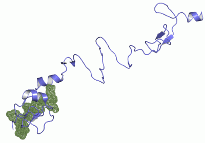
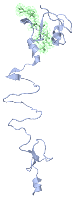
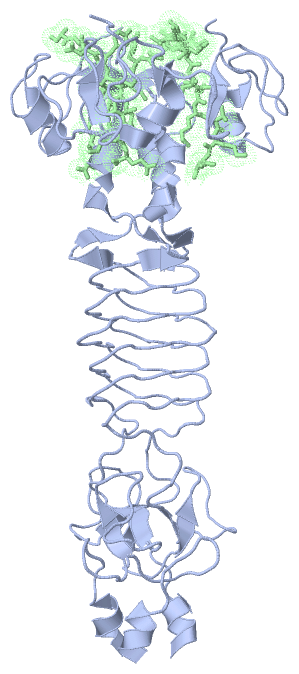
Asymmetric UnitChain A from PDB Type:PROTEIN Length:151 aligned with FIB12_BPT4 | P10930 from UniProtKB/Swiss-Prot Length:527 Alignment length:151 255 265 275 285 295 305 315 325 335 345 355 365 375 385 395 FIB12_BPT4 246 TGATLNGRGSTTSMRGVVKLTTTAGSQSGGDASSALAWNADVIQQRGGQIIYGTLRIEDTFTIANGGANITGTVRMTGGYIQGNRIVTQNEIDRTIPVGAIMMWAADSLPSDAWRFCHGGTVSASDCPLYASRIGTRYGGNPSNPGLPDMR 396 SCOP domains d1h6wa1 A:246-327 Middle part of short tail fibre protein gp12 d1h6w.2 A:328-396,B:518-527 SCOP domains CATH domains 1h6wA01 A:246-286 1h6wA02 A:287-329 1h6wA03 A:330-396 CATH domains Pfam domains ------------------------------------------------------------------------------------------------------------------------------------------------------- Pfam domains SAPs(SNPs) ------------------------------------------------------------------------------------------------------------------------------------------------------- SAPs(SNPs) PROSITE ------------------------------------------------------------------------------------------------------------------------------------------------------- PROSITE Transcript ------------------------------------------------------------------------------------------------------------------------------------------------------- Transcript 1h6w A 246 TGATLNGRGSTTSMRGVVKLTTTAGSQSGGDASSALAWNADVIHQRGGQTINGTLRINNTLTIASGGANITGTVNMTGGYIQGKRVVTQNEIDRTIPVGAIMMWAADSLPSDAWRFCHGGTVSASDCPLYASRIGTRYGGSSSNPGLPDMR 396 255 265 275 285 295 305 315 325 335 345 355 365 375 385 395 Chain B from PDB Type:PROTEIN Length:10 aligned with FIB12_BPT4 | P10930 from UniProtKB/Swiss-Prot Length:527 Alignment length:10 527 FIB12_BPT4 518 SLNYIIKVKE 527 SCOP domains d1h6w.2 SCOP domains CATH domains ---------- CATH domains Pfam domains ---------- Pfam domains SAPs(SNPs) ---------- SAPs(SNPs) PROSITE ---------- PROSITE Transcript ---------- Transcript 1h6w B 518 SLNYIIKVKE 527 527
|
||||||||||||||||||||
SCOP Domains (2, 2)
Asymmetric Unit
 |
CATH Domains (3, 3)
Asymmetric Unit
|
Pfam Domains (0, 0)| (no "Pfam Domain" information available for 1H6W) |
Gene Ontology (5, 5)|
Asymmetric Unit(hide GO term definitions) Chain A,B (FIB12_BPT4 | P10930)
|
||||||||||||||||||||||||||||||||||||||||||||||||
Interactive Views
|
||||||||||||||||||||||||||||||||||||||||||||||||||||||||||||||||||||||||||||||||||||||||||||||||||||||||||||||||||||||||||||||||||||||
Still Images
|
||||||||||||||||
Databases
|
||||||||||||||||||||||||||||||||||||||||||||||||||||||||||||||||||||||||||||||||||||||||||||||||||||||||||||||||||||||||||||||||||||||||||||||||||||||||||||||||
Analysis Tools
|
|||||||||||||||||||||||||||||||||||||||||||||||||||||||||||||
Entries Sharing at Least One Protein Chain (UniProt ID)
Related Entries Specified in the PDB File
|
|