| molecular function |
|---|
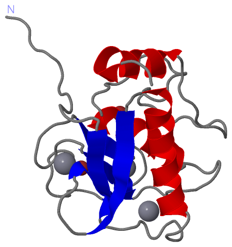
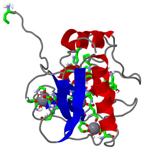
| | GO:0005509 | | calcium ion binding | | Interacting selectively and non-covalently with calcium ions (Ca2+). |
| | GO:0004175 | | endopeptidase activity | | Catalysis of the hydrolysis of internal, alpha-peptide bonds in a polypeptide chain. |
| | GO:0016787 | | hydrolase activity | | Catalysis of the hydrolysis of various bonds, e.g. C-O, C-N, C-C, phosphoric anhydride bonds, etc. Hydrolase is the systematic name for any enzyme of EC class 3. |
| | GO:0046872 | | metal ion binding | | Interacting selectively and non-covalently with any metal ion. |
| | GO:0004222 | | metalloendopeptidase activity | | Catalysis of the hydrolysis of internal, alpha-peptide bonds in a polypeptide chain by a mechanism in which water acts as a nucleophile, one or two metal ions hold the water molecule in place, and charged amino acid side chains are ligands for the metal ions. |
| | GO:0008237 | | metallopeptidase activity | | Catalysis of the hydrolysis of peptide bonds by a mechanism in which water acts as a nucleophile, one or two metal ions hold the water molecule in place, and charged amino acid side chains are ligands for the metal ions. |
| | GO:0008233 | | peptidase activity | | Catalysis of the hydrolysis of a peptide bond. A peptide bond is a covalent bond formed when the carbon atom from the carboxyl group of one amino acid shares electrons with the nitrogen atom from the amino group of a second amino acid. |
| | GO:0004252 | | serine-type endopeptidase activity | | Catalysis of the hydrolysis of internal, alpha-peptide bonds in a polypeptide chain by a catalytic mechanism that involves a catalytic triad consisting of a serine nucleophile that is activated by a proton relay involving an acidic residue (e.g. aspartate or glutamate) and a basic residue (usually histidine). |
| | GO:0008270 | | zinc ion binding | | Interacting selectively and non-covalently with zinc (Zn) ions. |
| biological process |
|---|
| | GO:0044267 | | cellular protein metabolic process | | The chemical reactions and pathways involving a specific protein, rather than of proteins in general, occurring at the level of an individual cell. Includes cellular protein modification. |
| | GO:0030574 | | collagen catabolic process | | The proteolytic chemical reactions and pathways resulting in the breakdown of collagen in the extracellular matrix, usually carried out by proteases secreted by nearby cells. |
| | GO:0022617 | | extracellular matrix disassembly | | A process that results in the breakdown of the extracellular matrix. |
| | GO:0050900 | | leukocyte migration | | The movement of a leukocyte within or between different tissues and organs of the body. |
| | GO:0032461 | | positive regulation of protein oligomerization | | Any process that activates or increases the frequency, rate or extent of protein oligomerization. |
| | GO:0006508 | | proteolysis | | The hydrolysis of proteins into smaller polypeptides and/or amino acids by cleavage of their peptide bonds. |
| | GO:0016032 | | viral process | | A multi-organism process in which a virus is a participant. The other participant is the host. Includes infection of a host cell, replication of the viral genome, and assembly of progeny virus particles. In some cases the viral genetic material may integrate into the host genome and only subsequently, under particular circumstances, 'complete' its life cycle. |
| cellular component |
|---|
| | GO:0031012 | | extracellular matrix | | A structure lying external to one or more cells, which provides structural support for cells or tissues. |
| | GO:0005576 | | extracellular region | | The space external to the outermost structure of a cell. For cells without external protective or external encapsulating structures this refers to space outside of the plasma membrane. This term covers the host cell environment outside an intracellular parasite. |
| | GO:0005578 | | proteinaceous extracellular matrix | | A layer consisting mainly of proteins (especially collagen) and glycosaminoglycans (mostly as proteoglycans) that forms a sheet underlying or overlying cells such as endothelial and epithelial cells. The proteins are secreted by cells in the vicinity. An example of this component is found in Mus musculus. |
 Description