Analysis of nucleic acid double helix geometry
| PDB code | | 3D2X (PDB summary) |
| Duplex length | | 77 base pairs |
Only the nucleic acid double helix part of the structure is analysed here. Small ligands, proteins, and overhanging ends are not taken into account. Information on the complete structure is available at the Image Library Entry page and at the Sequence, Chains, Units page.
| Strand 1 | 5' | G1 | G2 | G3 | A4 | C5 | C6 | A7 | G8 | G9 | G10 | G11 | U12 | G13 | C14 | U15 | U16 | G17 | U18 | U19 | C20 | A21 | C22 | A23 | G24 | G25 | C26 | U27 | G28 | A29 | G30 | A31 | A32 | A33 | G34 | U35 | C36 | C37 | C38 | U39 | U40 | U41 | G42 | A43 | A44 | C45 | C46 | U47 | G48 | A49 | A50 | C51 | A52 | G53 | G54 | G55 | U56 | A57 | A58 | U59 | G60 | C61 | C62 | U63 | G64 | C65 | G66 | C67 | A68 | G69 | G70 | G71 | A72 | G73 | U74 | G75 | U76 | C77 | 3' |
| Strand 2 | 3' | C77 | U76 | G75 | U74 | G73 | A72 | G71 | G70 | G69 | A68 | C67 | G66 | C65 | G64 | U63 | C62 | C61 | G60 | U59 | A58 | A57 | U56 | G55 | G54 | G53 | A52 | C51 | A50 | A49 | G48 | U47 | C46 | C45 | A44 | A43 | G42 | U41 | U40 | U39 | C38 | C37 | C36 | U35 | G34 | A33 | A32 | A31 | G30 | A29 | G28 | U27 | C26 | G25 | G24 | A23 | C22 | A21 | C20 | U19 | U18 | G17 | U16 | U15 | C14 | G13 | U12 | G11 | G10 | G9 | G8 | A7 | C6 | C5 | A4 | G3 | G2 | G1 | 5' |
CURVES analysis failed.
No curvilinear helical axis determined.
No helix parameters determined.
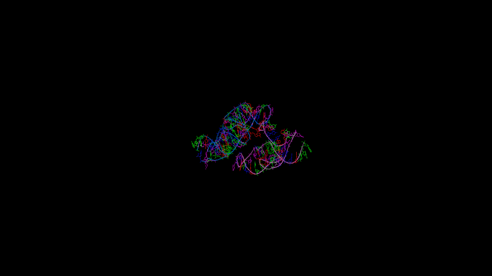
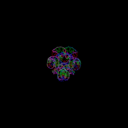
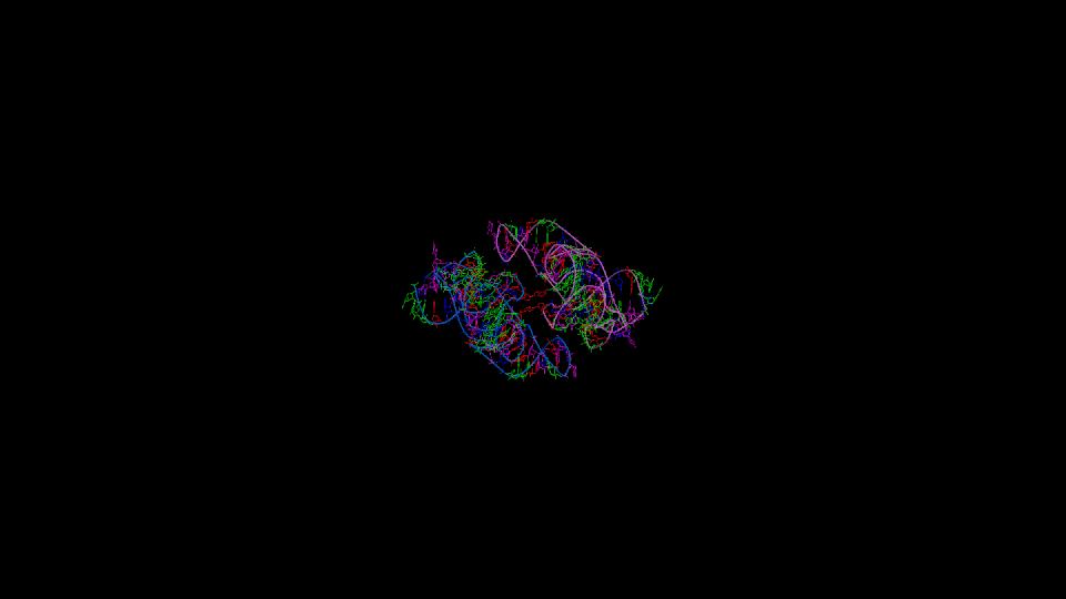
Figure 1
Three orthogonal views of the double helix (Help).
Residues are colored according to the nucleotide type (Help: Color codes).
The curvilinear helical axis (green) was calculated with
CURVES. The
double helix is oriented with respect to the principle axis of inertia
of the curvilinear helical axis (see Help for further explanations).
This drawing reveals immediately if there is any bending of the helical axis.
Further information
Full output from CURVES (helical parameters with respect to global and local axes)
Full output from FREEHELIX (helical parameters with respect to local axis, angles between normal vectors)
Chirality of ribose and phosphate atoms
Check the naming of phosphate and ribose substituents. Recommended for phosphate oxygens and for ribose hydrogens in NMR structures.


