Show PDB file:   
         Plain Text   HTML   (compressed file size)
QuickSearch:   
by PDB,NDB,UniProt,PROSITE Code or Search Term(s)  
(-)Asym.Unit - manually
(-)Asymmetric Unit
(-)Asym. Unit - sites
(-)Biological Unit 1
(-)Biol. Unit 1 - sites
collapse expand < >
Image Asym.Unit - manually
Asym.Unit - manually  (Jmol Viewer)
Image Asymmetric Unit
Asymmetric Unit  (Jmol Viewer)
Image Asym. Unit - sites
Asym. Unit - sites  (Jmol Viewer)
Image Biological Unit 1
Biological Unit 1  (Jmol Viewer)
Image Biol. Unit 1 - sites
Biol. Unit 1 - sites  (Jmol Viewer)

(-) Description

Title :  HUMAN PHENYLALANINE HYDROXYLASE CATALYTIC DOMAIN DIMER WITH BOUND L-DOPA (3,4-DIHYDROXYPHENYLALANINE) INHIBITOR
 
Authors :  H. Erlandsen, T. Flatmark, R. C. Stevens
Date :  20 Aug 98  (Deposition) - 27 Apr 99  (Release) - 31 Aug 11  (Revision)
Method :  X-RAY DIFFRACTION
Resolution :  2.15
Chains :  Asym. Unit :  A
Biol. Unit 1:  A  (2x)
Keywords :  Non-Heme Iron-Containing Monooxygenase, Oxidoreductase, Monooxygenase (Keyword Search: [Gene Ontology, PubMed, Web (Google))
 
Reference :  H. Erlandsen, T. Flatmark, R. C. Stevens, E. Hough
Crystallographic Analysis Of The Human Phenylalanine Hydroxylase Catalytic Domain With Bound Catechol Inhibitors At 2. 0 A Resolution.
Biochemistry V. 37 15638 1998
PubMed-ID: 9843368  |  Reference-DOI: 10.1021/BI9815290

(-) Compounds

Molecule 1 - PHENYLALANINE 4-MONOOXYGENASE
    ChainsA
    EC Number1.14.16.1
    EngineeredYES
    Expression SystemESCHERICHIA COLI
    Expression System Taxid562
    FragmentCATALYTIC DOMAIN
    OrganLIVER
    Organism CommonHUMAN
    Organism ScientificHOMO SAPIENS
    Organism Taxid9606
    Other DetailsTRUNCATED FORM, DELTA NH 1-102, DELTA COOH 428-452

 Structural Features

(-) Chains, Units

  1
Asymmetric Unit A
Biological Unit 1 (2x)A

Summary Information (see also Sequences/Alignments below)
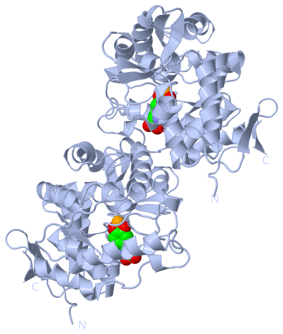
(-) Ligands, Modified Residues, Ions  (2, 2)

Asymmetric Unit (2, 2)
No.NameCountTypeFull Name
1DAH1Ligand/Ion3,4-DIHYDROXYPHENYLALANINE
2FE1Ligand/IonFE (III) ION
Biological Unit 1 (1, 2)
No.NameCountTypeFull Name
1DAH2Ligand/Ion3,4-DIHYDROXYPHENYLALANINE
2FE-1Ligand/IonFE (III) ION

(-) Sites  (3, 3)

Asymmetric Unit (3, 3)
No.NameEvidenceResiduesDescription
1AC1SOFTWAREHIS A:285 , HIS A:290 , GLU A:330 , DAH A:600 , HOH A:649BINDING SITE FOR RESIDUE FE A 425
2AC2SOFTWARELYS A:199 , LEU A:248 , HIS A:285 , HIS A:290 , TYR A:325 , GLU A:330 , FE A:425 , HOH A:649BINDING SITE FOR RESIDUE DAH A 600
3NULUNKNOWNHIS A:285 , HIS A:290 , GLU A:330 , HOH A:649 , DAH A:600IRON BINDING LIGANDS

(-) SS Bonds  (0, 0)

(no "SS Bond" information available for 6PAH)

(-) Cis Peptide Bonds  (0, 0)

(no "Cis Peptide Bond" information available for 6PAH)

 Sequence-Structure Mapping

(-) SAPs(SNPs)/Variants  (0, 0)

(no "SAP(SNP)/Variant" information available for 6PAH)

(-) PROSITE Motifs  (0, 0)

(no "PROSITE Motif" information available for 6PAH)

(-) Exons   (0, 0)

(no "Exon" information available for 6PAH)

(-) Sequences/Alignments

Asymmetric Unit
   Reformat: Number of residues per line =  ('0' or empty: single-line sequence representation)
  Number of residues per labelling interval =   
  UniProt sequence: complete  aligned part    
   Show mapping: SCOP domains CATH domains Pfam domains Secondary structure (by author)
SAPs(SNPs) PROSITE motifs Exons
(details for a mapped element are shown in a popup box when the mouse pointer rests over it)
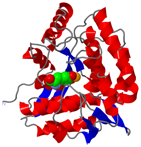
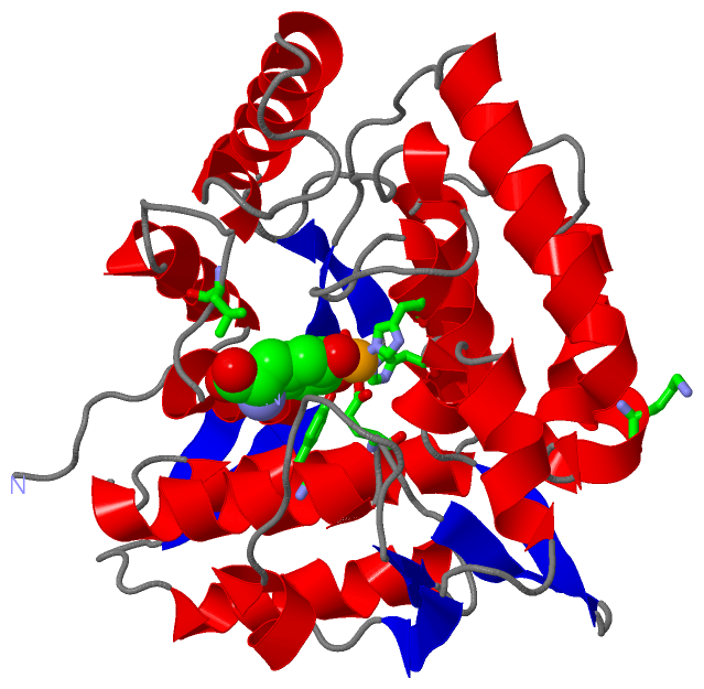
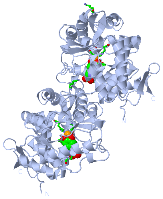
Chain A from PDB  Type:PROTEIN  Length:308
                                                                                                                                                                                                                                                                                                                                                    
               SCOP domains d6paha_ A: Phenylalanine hydroxylase, PAH                                                                                                                                                                                                                                                                            SCOP domains
               CATH domains 6pahA00 A:117-424 Phenylalanine Hydroxylase                                                                                                                                                                                                                                                                          CATH domains
               Pfam domains -------------------------------------------------------------------------------------------------------------------------------------------------------------------------------------------------------------------------------------------------------------------------------------------------------------------- Pfam domains
         Sec.struct. author ........hhhhhhhhhh.....hhh.........hhhhhhhhhhhhhhhh.............hhhhhhhhhhhhhhhhhhhhh..hhhhhhhhhhhhh..........hhhhhhhhhhhh..eeee......hhhhhhhhhh.eeee......hhh........hhhhhhh.hhhhh.hhhhhhhhhhhhhh....hhhhhhhhhhhhh.....eeee..eeee..hhhh..hhhhhhhh....eeee..hhhhhh..........eeee...hhhhhhhhhhhhhh.....eeeee....eeeee Sec.struct. author
                 SAPs(SNPs) -------------------------------------------------------------------------------------------------------------------------------------------------------------------------------------------------------------------------------------------------------------------------------------------------------------------- SAPs(SNPs)
                    PROSITE -------------------------------------------------------------------------------------------------------------------------------------------------------------------------------------------------------------------------------------------------------------------------------------------------------------------- PROSITE
                 Transcript -------------------------------------------------------------------------------------------------------------------------------------------------------------------------------------------------------------------------------------------------------------------------------------------------------------------- Transcript
                 6pah A 117 TVPWFPRTIQELDRFANQILSYGAELDADHPGFKDPVYRARRKQFADIAYNYRHGQPIPRVEYMEEEKKTWGTVFKTLKSLYKTHACYEYNHIFPLLEKYCGFHEDNIPQLEDVSQFLQTCTGFRLRPVAGLLSSRDFLGGLAFRVFHCTQYIRHGSKPMYTPEPDICHELLGHVPLFSDRSFAQFSQEIGLASLGAPDEYIEKLATIYWFTVEFGLCKQGDSIKAYGAGLLSSFGELQYCLSEKPKLLPLELEKTAIQNYTVTEFQPLYYVAESFNDAKEKVRNFAATIPRPFSVRYDPYTQRIEVL 424
                                   126       136       146       156       166       176       186       196       206       216       226       236       246       256       266       276       286       296       306       316       326       336       346       356       366       376       386       396       406       416        

   Legend:   → Mismatch (orange background)
  - → Gap (green background, '-', border residues have a numbering label)
    → Modified Residue (blue background, lower-case, 'x' indicates undefined single-letter code, labelled with number + name)
  x → Chemical Group (purple background, 'x', labelled with number + name, e.g. ACE or NH2)
  extra numbering lines below/above indicate numbering irregularities and modified residue names etc., number ends below/above '|'

 Classification and Annotation

(-) SCOP Domains  (1, 1)

Asymmetric Unit

(-) CATH Domains  (1, 1)

Asymmetric Unit

(-) Pfam Domains  (0, 0)

(no "Pfam Domain" information available for 6PAH)

(-) Gene Ontology  (24, 24)

Asymmetric Unit(hide GO term definitions)

 Visualization

(-) Interactive Views

Asymmetric Unit
  Complete Structure
    Jena3D(integrated viewing of ligand, site, SAP, PROSITE, SCOP information)
    WebMol | AstexViewer[tm]@PDBe
(Java Applets, require no local installation except for Java; loading may be slow)
    STRAP
(Java WebStart application, automatic local installation, requires Java; full application with system access!)
    RasMol
(require local installation)
    Molscript (VRML)
(requires installation of a VRML viewer; select preferred view via VRML and generate a mono or stereo PDF format file)
 
  Ligands, Modified Residues, Ions
    DAH  [ RasMol | Jena3D ]  +environment [ RasMol | Jena3D ]
    FE  [ RasMol | Jena3D ]  +environment [ RasMol | Jena3D ]
 
  Sites
    AC1  [ RasMol ]  +environment [ RasMol ]
    AC2  [ RasMol ]  +environment [ RasMol ]
    NUL  [ RasMol ]  +environment [ RasMol ]
 
  Cis Peptide Bonds
(no "Cis Peptide Bonds" information available for 6pah)
 
Biological Unit
  Complete Structure
    Biological Unit 1  [ Jena3D ]

(-) Still Images

Jmol
  protein: cartoon or spacefill or dots and stick; nucleic acid: cartoon and stick; ligands: spacefill; active site: stick
Molscript
  protein, nucleic acid: cartoon; ligands: spacefill; active site: ball and stick

 Databases and Analysis Tools

(-) Databases

Access by PDB/NDB ID
  6pah
    Family and Domain InformationProDom | SYSTERS
    General Structural InformationGlycoscienceDB | MMDB | NDB | OCA | PDB | PDBe | PDBj | PDBsum | PDBWiki | PQS | PROTEOPEDIA
    Orientation in MembranesOPM
    Protein SurfaceSURFACE
    Secondary StructureDSSP (structure derived) | HSSP (homology derived)
    Structural GenomicsGeneCensus
    Structural NeighboursCE | VAST
    Structure ClassificationCATH | Dali | SCOP
    Validation and Original DataBMRB Data View | BMRB Restraints Grid | EDS | PROCHECK | RECOORD | WHAT_CHECK
 
Access by UniProt ID/Accession number
  PH4H_HUMAN | P00439
    Comparative Protein Structure ModelsModBase
    Genomic InformationEnsembl
    Protein-protein InteractionDIP
    Sequence, Family and Domain InformationInterPro | Pfam | SMART | UniProtKB/SwissProt
 
Access by Enzyme Classificator   (EC Number)
  1.14.16.1
    General Enzyme InformationBRENDA | EC-PDB | Enzyme | IntEnz
    PathwayKEGG | MetaCyc
 
Access by Disease Identifier   (MIM ID)
  (no 'MIM ID' available)
    Disease InformationOMIM
 
Access by GenAge ID
  (no 'GenAge ID' available)
    Age Related InformationGenAge

(-) Analysis Tools

Access by PDB/NDB ID
    Domain InformationXDom
    Interatomic Contacts of Structural UnitsCSU
    Ligand-protein ContactsLPC
    Protein CavitiescastP
    Sequence and Secondary StructurePDBCartoon
    Structure AlignmentSTRAP(Java WebStart application, automatic local installation, requires Java; full application with system access!)
    Structure and Sequence BrowserSTING
 
Access by UniProt ID/Accession number
  PH4H_HUMAN | P00439
    Protein Disorder PredictionDisEMBL | FoldIndex | GLOBPLOT (for more information see DisProt)

 Related Entries

(-) Entries Sharing at Least One Protein Chain (UniProt ID)

UniProtKB/Swiss-Prot
        PH4H_HUMAN | P004391dmw 1j8t 1j8u 1kw0 1lrm 1mmk 1mmt 1pah 1tdw 1tg2 2pah 3pah 4anp 4pah 5fii 5pah

(-) Related Entries Specified in the PDB File

(no "Related Entries Specified in the PDB File" available for 6PAH)