|
|
|
|
 Description
|
|
Compounds
|
||||||||||||||||||||||||||||||||||||||||
Chains, Units
Summary Information (see also Sequences/Alignments below) |
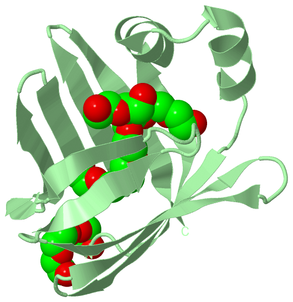
Ligands, Modified Residues, Ions (4, 4)
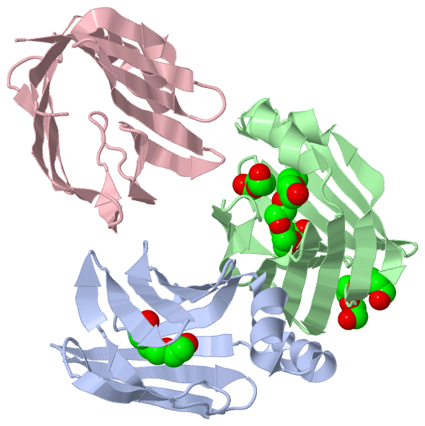
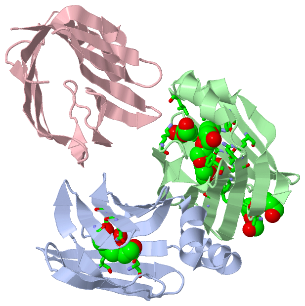
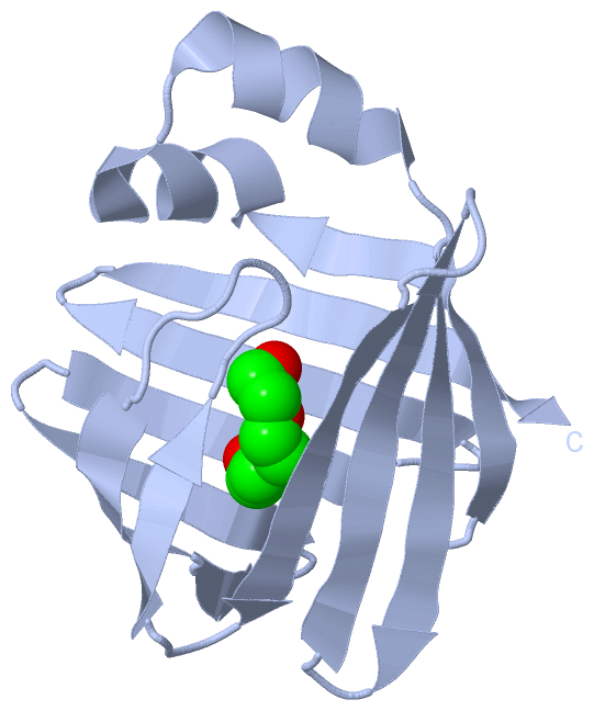
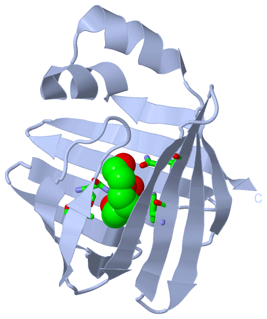
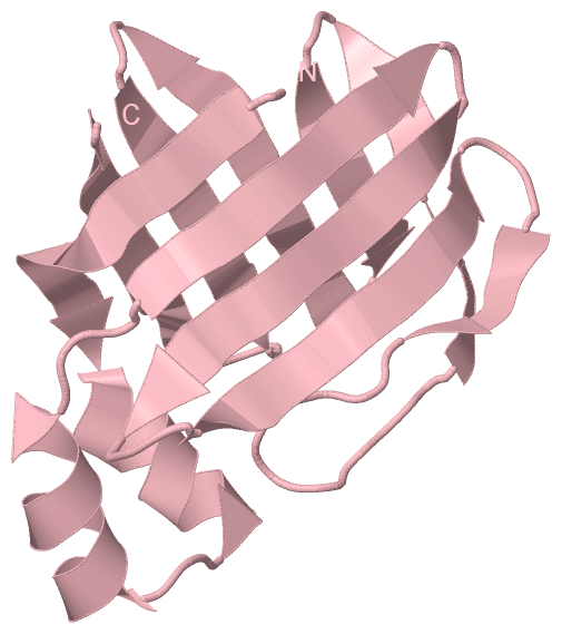
Asymmetric Unit (4, 4)
|
Sites (4, 4)
Asymmetric Unit (4, 4)
|
SS Bonds (0, 0)| (no "SS Bond" information available for 5L8I) |
Cis Peptide Bonds (0, 0)| (no "Cis Peptide Bond" information available for 5L8I) |
SAPs(SNPs)/Variants (0, 0)| (no "SAP(SNP)/Variant" information available for 5L8I) |
PROSITE Motifs (0, 0)| (no "PROSITE Motif" information available for 5L8I) |
Exons (0, 0)| (no "Exon" information available for 5L8I) |
Sequences/Alignments
Asymmetric Unit
Chain A from PDB Type:PROTEIN Length:125
SCOP domains ----------------------------------------------------------------------------------------------------------------------------- SCOP domains
CATH domains ----------------------------------------------------------------------------------------------------------------------------- CATH domains
Pfam domains ----------------------------------------------------------------------------------------------------------------------------- Pfam domains
SAPs(SNPs) ----------------------------------------------------------------------------------------------------------------------------- SAPs(SNPs)
PROSITE ----------------------------------------------------------------------------------------------------------------------------- PROSITE
Transcript ----------------------------------------------------------------------------------------------------------------------------- Transcript
5l8i A 3 FTGKFEMESEKNYDEFMKLLGISSDVIEKARNFKIVTEVQQDGQDFTWSQHYSGGHTMTNKFTVGKESNIQTMGGKTFKATVQMEGGKLVVNFPNYHQTSEIVGDKLVEVSTIGGVTYERVSKRL 127
12 22 32 42 52 62 72 82 92 102 112 122
Chain B from PDB Type:PROTEIN Length:126
SCOP domains ------------------------------------------------------------------------------------------------------------------------------ SCOP domains
CATH domains ------------------------------------------------------------------------------------------------------------------------------ CATH domains
Pfam domains ------------------------------------------------------------------------------------------------------------------------------ Pfam domains
SAPs(SNPs) ------------------------------------------------------------------------------------------------------------------------------ SAPs(SNPs)
PROSITE ------------------------------------------------------------------------------------------------------------------------------ PROSITE
Transcript ------------------------------------------------------------------------------------------------------------------------------ Transcript
5l8i B 2 AFTGKFEMESEKNYDEFMKLLGISSDVIEKARNFKIVTEVQQDGQDFTWSQHYSGGHTMTNKFTVGKESNIQTMGGKTFKATVQMEGGKLVVNFPNYHQTSEIVGDKLVEVSTIGGVTYERVSKRL 127
11 21 31 41 51 61 71 81 91 101 111 121
Chain C from PDB Type:PROTEIN Length:125
SCOP domains ----------------------------------------------------------------------------------------------------------------------------- SCOP domains
CATH domains ----------------------------------------------------------------------------------------------------------------------------- CATH domains
Pfam domains ----------------------------------------------------------------------------------------------------------------------------- Pfam domains
SAPs(SNPs) ----------------------------------------------------------------------------------------------------------------------------- SAPs(SNPs)
PROSITE ----------------------------------------------------------------------------------------------------------------------------- PROSITE
Transcript ----------------------------------------------------------------------------------------------------------------------------- Transcript
5l8i C 3 FTGKFEMESEKNYDEFMKLLGISSDVIEKARNFKIVTEVQQDGQDFTWSQHYSGGHTMTNKFTVGKESNIQTMGGKTFKATVQMEGGKLVVNFPNYHQTSEIVGDKLVEVSTIGGVTYERVSKRL 127
12 22 32 42 52 62 72 82 92 102 112 122
|
||||||||||||||||||||
SCOP Domains (0, 0)| (no "SCOP Domain" information available for 5L8I) |
CATH Domains (0, 0)| (no "CATH Domain" information available for 5L8I) |
Pfam Domains (0, 0)| (no "Pfam Domain" information available for 5L8I) |
Gene Ontology (13, 13)|
Asymmetric Unit(hide GO term definitions) |
Interactive Views
|
||||||||||||||||||||||||||||||||||||||||||||||||||||||||||||||||||||||||||||||||||||||||||||||||||||||||||||||||||||||||||||||||||||||||||||||||||||||||||||||||||||||||||||||||||||||||||||
Still Images
|
||||||||||||||||
Databases
|
||||||||||||||||||||||||||||||||||||||||||||||||||||||||||||||||||||||||||||||||||||||||||||||||||||||||||||||||||||||||||||||||||||||||||||||||||||||||||||||||
Analysis Tools
|
|||||||||||||||||||||||||||||||||||||||||||||||||||||||||||||
Entries Sharing at Least One Protein Chain (UniProt ID)
Related Entries Specified in the PDB File
|
|