Show PDB file:   
         Plain Text   HTML   (compressed file size)
QuickSearch:   
by PDB,NDB,UniProt,PROSITE Code or Search Term(s)  
(-)Asym./Biol. Unit
(-)Asym./Biol. Unit - sites
collapse expand < >
Image Asym./Biol. Unit
Asym./Biol. Unit  (Jmol Viewer)
Image Asym./Biol. Unit - sites
Asym./Biol. Unit - sites  (Jmol Viewer)

(-) Description

Title :  CHOLERA TOXIN CLASSICAL B-PENTAMER IN COMPLEX WITH A LEWIS-Y
 
Authors :  J. E. Heggelund, D. Burschowsky, U. Krengel
Date :  04 Nov 15  (Deposition) - 30 Mar 16  (Release) - 11 May 16  (Revision)
Method :  X-RAY DIFFRACTION
Resolution :  1.40
Chains :  Asym./Biol. Unit :  A,B,C,D,E
Keywords :  Cholera Toxin B-Pentamer, A Lewis-Y, Complex, Blood Group Oligosaccharide/Antigen, Toxin (Keyword Search: [Gene Ontology, PubMed, Web (Google))
 
Reference :  J. E. Heggelund, D. Burschowsky, V. A. Bjrnestad, V. Hodnik, G. Anderluh, U. Krengel
High-Resolution Crystal Structures Elucidate The Molecular Basis Of Cholera Blood Group Dependence.
Plos Pathog. V. 12 05567 2016
PubMed-ID: 27082955  |  Reference-DOI: 10.1371/JOURNAL.PPAT.1005567

(-) Compounds

Molecule 1 - CHOLERA ENTEROTOXIN B SUBUNIT
    ChainsA, B, C, D, E
    EngineeredYES
    Expression SystemESCHERICHIA COLI BL21(DE3)
    Expression System Taxid469008
    GeneCTXB
    Organism ScientificVIBRIO CHOLERAE O1
    Organism Taxid127906
    SynonymCHOLERA TOXIN B-PENTAMER, CLASSICAL VARIANT,CHOLERA ENTEROTOXIN SUBUNIT B,CHOLERA TOXIN B PROTEIN (CTB),CHOLERA TOXIN B SUBUNIT,CHOLERA TOXIN BETA SUBUNIT,CHOLERA TOXIN SUBUNIT B,CHOLERAE TOXIN B SUBUNIT,CTXB

 Structural Features

(-) Chains, Units

  12345
Asymmetric/Biological Unit ABCDE

Summary Information (see also Sequences/Alignments below)

(-) Ligands, Modified Residues, Ions  (9, 31)

Asymmetric/Biological Unit (9, 31)
No.NameCountTypeFull Name
1A2G4Ligand/IonN-ACETYL-2-DEOXY-2-AMINO-GALACTOSE
2FUC8Ligand/IonALPHA-L-FUCOSE
3GAL7Ligand/IonBETA-D-GALACTOSE
4GLA1Ligand/IonALPHA D-GALACTOSE
5MES1Ligand/Ion2-(N-MORPHOLINO)-ETHANESULFONIC ACID
6NA1Ligand/IonSODIUM ION
7NDG4Ligand/Ion2-(ACETYLAMINO)-2-DEOXY-A-D-GLUCOPYRANOSE
8PEG2Ligand/IonDI(HYDROXYETHYL)ETHER
9PGE3Ligand/IonTRIETHYLENE GLYCOL

(-) Sites  (14, 14)

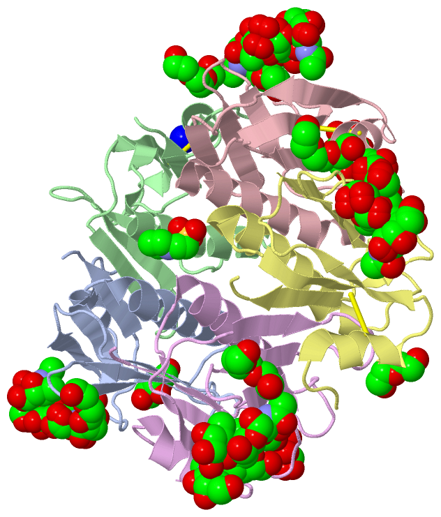
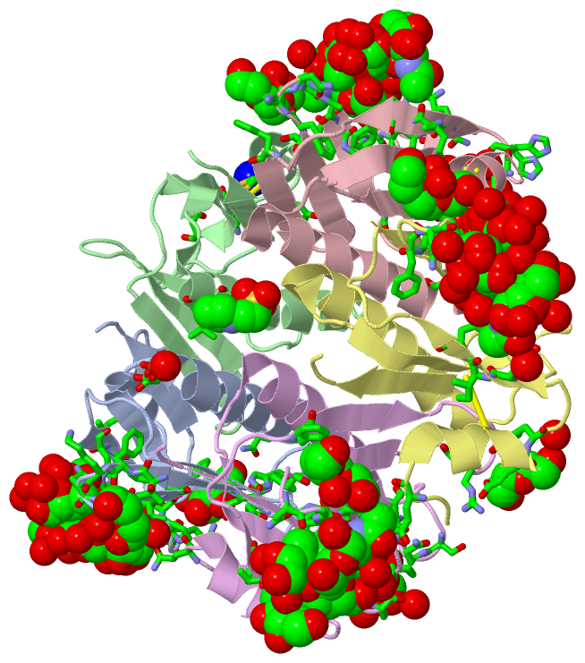
Asymmetric Unit (14, 14)
No.NameEvidenceResiduesDescription
01AC1SOFTWARETHR A:41 , PHE A:42 , LYS A:43 , NDG A:203 , HOH A:383 , HOH A:385binding site for residue PEG A 201
02AC2SOFTWAREGLU A:51 , GLN A:56 , HIS A:57 , GLN A:61 , TRP A:88 , ASN A:90 , LYS A:91 , HOH A:361binding site for residue GAL A 202
03AC3SOFTWAREGLU A:79 , HOH A:399 , CYS B:9 , TYR B:12 , THR B:15 , HOH B:366binding site for residue NA B 201
04AC4SOFTWAREASN B:14 , ILE B:74 , LEU B:77 , ASN B:89binding site for residue MES C 201
05AC5SOFTWAREPHE C:25 , THR C:41 , PHE C:42 , LYS C:43 , GLY C:45 , NDG C:204 , HOH C:387binding site for residue PGE C 202
06AC6SOFTWAREGLU C:51 , GLN C:56 , HIS C:57 , GLN C:61 , TRP C:88 , ASN C:90 , LYS C:91 , HOH C:304 , HOH C:306 , HOH C:362 , HOH C:388 , GLN D:16 , HOH D:376binding site for residue GAL C 203
07AC7SOFTWAREGLU D:11 , TYR D:12 , HOH D:395 , ARG E:35binding site for residue PEG D 201
08AC8SOFTWARETHR D:41 , PHE D:42 , NDG D:203 , HOH D:365 , HOH D:380binding site for residue PGE D 202
09AC9SOFTWAREPHE E:25 , THR E:41 , PHE E:42 , NDG E:204 , HOH E:391binding site for residue PGE E 201
10AD1SOFTWAREGLN A:16 , ASN A:89 , HOH A:311 , HOH A:319 , GLU E:51 , GLN E:56 , HIS E:57 , GLN E:61 , TRP E:88 , ASN E:90 , LYS E:91 , HOH E:303 , HOH E:361 , HOH E:378binding site for residue GAL E 202
11AD2SOFTWAREGLN A:16 , ILE A:17 , HIS A:18 , GLY A:33 , LYS A:34 , ASN A:44 , GLY A:45 , ALA A:46 , THR A:47 , PHE A:48 , PRO A:93 , HIS A:94 , PEG A:201 , HOH A:303 , HOH A:304 , HOH A:308 , HOH A:309 , HOH A:311 , HOH A:312 , HOH A:318 , HOH A:320 , HOH A:322 , HOH A:324 , HOH A:328 , HOH A:333 , HOH A:336 , HOH A:350 , HOH A:369 , HOH A:387 , HOH A:392 , GLN E:3 , SER E:55 , GLN E:56 , HIS E:57 , ILE E:58 , HOH E:341binding site for Poly-Saccharide residues NDG A 203 through FUC A 207
12AD3SOFTWAREGLN A:16 , ILE A:17 , HIS A:18 , GLY A:33 , LYS A:34 , ASN A:44 , GLY A:45 , ALA A:46 , THR A:47 , PHE A:48 , PRO A:93 , HIS A:94 , PEG A:201 , HOH A:303 , HOH A:304 , HOH A:308 , HOH A:309 , HOH A:311 , HOH A:312 , HOH A:318 , HOH A:320 , HOH A:322 , HOH A:324 , HOH A:328 , HOH A:333 , HOH A:336 , HOH A:350 , HOH A:369 , HOH A:387 , HOH A:392 , GLN B:3 , GLN C:16 , ILE C:17 , HIS C:18 , ASN C:44 , GLY C:45 , ALA C:46 , THR C:47 , PHE C:48 , GLU C:51 , GLN C:56 , HIS C:57 , GLN C:61 , TRP C:88 , ASN C:90 , LYS C:91 , THR C:92 , PRO C:93 , HIS C:94 , PGE C:202 , HOH C:304 , HOH C:305 , HOH C:306 , HOH C:313 , HOH C:315 , HOH C:318 , HOH C:362 , HOH C:388 , HOH C:397 , GLN D:16 , HOH D:376 , GLN E:3 , SER E:55 , GLN E:56 , HIS E:57 , ILE E:58 , A2G E:206 , HOH E:341binding site for Poly-Saccharide residues NDG C 204 through FUC C 208
13AD4SOFTWAREGLN B:3 , GLN C:3 , TYR C:12 , HIS C:13 , GLN C:16 , ILE C:17 , HIS C:18 , ASN C:44 , GLY C:45 , ALA C:46 , THR C:47 , GLU C:51 , SER C:55 , GLN C:56 , HIS C:57 , ILE C:58 , GLN C:61 , TRP C:88 , ASN C:90 , LYS C:91 , HIS C:94 , PGE C:202 , FUC C:208 , HOH C:304 , HOH C:305 , HOH C:306 , HOH C:313 , HOH C:315 , HOH C:324 , HOH C:362 , HOH C:388 , HOH C:397 , GLN D:16 , ILE D:17 , HIS D:18 , GLY D:33 , LYS D:34 , ASN D:44 , GLY D:45 , ALA D:46 , THR D:47 , PHE D:48 , PRO D:93 , HIS D:94 , PGE D:202 , HOH D:302 , HOH D:303 , HOH D:305 , HOH D:306 , HOH D:309 , HOH D:313 , HOH D:314 , HOH D:316 , HOH D:332 , HOH D:346 , HOH D:374 , HOH D:376 , HOH D:379 , HOH D:390 , HOH D:391 , A2G E:206binding site for Poly-Saccharide residues NDG D 203 through FUC D 207
14AD5SOFTWAREGLN C:3 , TYR C:12 , HIS C:13 , SER C:55 , GLN C:56 , HIS C:57 , ILE C:58 , A2G C:206 , HOH C:304 , HOH C:324 , HOH C:362 , GLN D:3 , GLN D:16 , ILE D:17 , HIS D:18 , GLY D:33 , LYS D:34 , ASN D:44 , GLY D:45 , ALA D:46 , THR D:47 , PHE D:48 , PRO D:93 , HIS D:94 , PGE D:202 , HOH D:302 , HOH D:303 , HOH D:305 , HOH D:306 , HOH D:309 , HOH D:313 , HOH D:314 , HOH D:316 , HOH D:332 , HOH D:346 , HOH D:374 , HOH D:379 , HOH D:390 , HOH D:391 , GLN E:16 , ILE E:17 , HIS E:18 , ASN E:44 , GLY E:45 , ALA E:46 , THR E:47 , PHE E:48 , PRO E:93 , HIS E:94 , PGE E:201 , HOH E:304 , HOH E:308 , HOH E:326 , HOH E:388binding site for Poly-Saccharide residues NDG E 203 through FUC E 207

(-) SS Bonds  (5, 5)

Asymmetric/Biological Unit
No.Residues
1A:9 -A:86
2B:9 -B:86
3C:9 -C:86
4D:9 -D:86
5E:9 -E:86

(-) Cis Peptide Bonds  (5, 6)

Asymmetric/Biological Unit
No.Residues
1Thr A:92 -Pro A:93
2Thr B:92 -Pro B:93
3Thr C:92 -Pro C:93
4Thr D:92 -Pro D:93
5Thr E:92 -Pro E:93

 Sequence-Structure Mapping

(-) SAPs(SNPs)/Variants  (0, 0)

(no "SAP(SNP)/Variant" information available for 5ELD)

(-) PROSITE Motifs  (0, 0)

(no "PROSITE Motif" information available for 5ELD)

(-) Exons   (0, 0)

(no "Exon" information available for 5ELD)

(-) Sequences/Alignments

Asymmetric/Biological Unit
   Reformat: Number of residues per line =  ('0' or empty: single-line sequence representation)
  Number of residues per labelling interval =   
  UniProt sequence: complete  aligned part    
   Show mapping: SCOP domains CATH domains Pfam domains Secondary structure (by author)
SAPs(SNPs) PROSITE motifs Exons
(details for a mapped element are shown in a popup box when the mouse pointer rests over it)
Chain A from PDB  Type:PROTEIN  Length:103
                                                                                                                                       
               SCOP domains ------------------------------------------------------------------------------------------------------- SCOP domains
               CATH domains ------------------------------------------------------------------------------------------------------- CATH domains
               Pfam domains ------------------------------------------------------------------------------------------------------- Pfam domains
         Sec.struct. author ...hhhhhhh....eeeee......eeee........eeee.....eeee.........hhhhhhhhhhhhhhhhhhhh..eeeeeee.........eeee.. Sec.struct. author
                 SAPs(SNPs) ------------------------------------------------------------------------------------------------------- SAPs(SNPs)
                    PROSITE ------------------------------------------------------------------------------------------------------- PROSITE
                 Transcript ------------------------------------------------------------------------------------------------------- Transcript
                 5eld A   1 TPQNITDLCAEYHNTQIHTLNDKIFSYTESLAGKREMAIITFKNGATFQVEVPGSQHIDSQKKAIERMKDTLRIAYLTEAKVEKLCVWNNKTPHAIAAISMAN 103
                                    10        20        30        40        50        60        70        80        90       100   

Chain B from PDB  Type:PROTEIN  Length:103
                                                                                                                                       
               SCOP domains ------------------------------------------------------------------------------------------------------- SCOP domains
               CATH domains ------------------------------------------------------------------------------------------------------- CATH domains
               Pfam domains ------------------------------------------------------------------------------------------------------- Pfam domains
         Sec.struct. author ...hhhhhhh....eeeee......eeeee......eeeee.....eeee.........hhhhhhhhhhhhhhhhhhhh..eeeeeee.........eeee.. Sec.struct. author
                 SAPs(SNPs) ------------------------------------------------------------------------------------------------------- SAPs(SNPs)
                    PROSITE ------------------------------------------------------------------------------------------------------- PROSITE
                 Transcript ------------------------------------------------------------------------------------------------------- Transcript
                 5eld B   1 TPQNITDLCAEYHNTQIHTLNDKIFSYTESLAGKREMAIITFKNGATFQVEVPGSQHIDSQKKAIERMKDTLRIAYLTEAKVEKLCVWNNKTPHAIAAISMAN 103
                                    10        20        30        40        50        60        70        80        90       100   

Chain C from PDB  Type:PROTEIN  Length:103
                                                                                                                                       
               SCOP domains ------------------------------------------------------------------------------------------------------- SCOP domains
               CATH domains ------------------------------------------------------------------------------------------------------- CATH domains
               Pfam domains ------------------------------------------------------------------------------------------------------- Pfam domains
         Sec.struct. author ...hhhhhhh....eeeee......eeeee......eeeee.....eeee.......hhhhhhhhhhhhhhhhhhhhhh..eeeeeee.........eeee.. Sec.struct. author
                 SAPs(SNPs) ------------------------------------------------------------------------------------------------------- SAPs(SNPs)
                    PROSITE ------------------------------------------------------------------------------------------------------- PROSITE
                 Transcript ------------------------------------------------------------------------------------------------------- Transcript
                 5eld C   1 TPQNITDLCAEYHNTQIHTLNDKIFSYTESLAGKREMAIITFKNGATFQVEVPGSQHIDSQKKAIERMKDTLRIAYLTEAKVEKLCVWNNKTPHAIAAISMAN 103
                                    10        20        30        40        50        60        70        80        90       100   

Chain D from PDB  Type:PROTEIN  Length:103
                                                                                                                                       
               SCOP domains ------------------------------------------------------------------------------------------------------- SCOP domains
               CATH domains ------------------------------------------------------------------------------------------------------- CATH domains
               Pfam domains ------------------------------------------------------------------------------------------------------- Pfam domains
         Sec.struct. author ...hhhhhhhh...eeeee......eeeee......eeeee.....eeee.........hhhhhhhhhhhhhhhhhhhh..eeeeeee.........eeee.. Sec.struct. author
                 SAPs(SNPs) ------------------------------------------------------------------------------------------------------- SAPs(SNPs)
                    PROSITE ------------------------------------------------------------------------------------------------------- PROSITE
                 Transcript ------------------------------------------------------------------------------------------------------- Transcript
                 5eld D   1 TPQNITDLCAEYHNTQIHTLNDKIFSYTESLAGKREMAIITFKNGATFQVEVPGSQHIDSQKKAIERMKDTLRIAYLTEAKVEKLCVWNNKTPHAIAAISMAN 103
                                    10        20        30        40        50        60        70        80        90       100   

Chain E from PDB  Type:PROTEIN  Length:103
                                                                                                                                       
               SCOP domains ------------------------------------------------------------------------------------------------------- SCOP domains
               CATH domains ------------------------------------------------------------------------------------------------------- CATH domains
               Pfam domains ------------------------------------------------------------------------------------------------------- Pfam domains
         Sec.struct. author ...hhhhhhh....eeeee......eeee........eeee.....eeee.......hhhhhhhhhhhhhhhhhhhhh...eeeeeee.........eeee.. Sec.struct. author
                 SAPs(SNPs) ------------------------------------------------------------------------------------------------------- SAPs(SNPs)
                    PROSITE ------------------------------------------------------------------------------------------------------- PROSITE
                 Transcript ------------------------------------------------------------------------------------------------------- Transcript
                 5eld E   1 TPQNITDLCAEYHNTQIHTLNDKIFSYTESLAGKREMAIITFKNGATFQVEVPGSQHIDSQKKAIERMKDTLRIAYLTEAKVEKLCVWNNKTPHAIAAISMAN 103
                                    10        20        30        40        50        60        70        80        90       100   

   Legend:   → Mismatch (orange background)
  - → Gap (green background, '-', border residues have a numbering label)
    → Modified Residue (blue background, lower-case, 'x' indicates undefined single-letter code, labelled with number + name)
  x → Chemical Group (purple background, 'x', labelled with number + name, e.g. ACE or NH2)
  extra numbering lines below/above indicate numbering irregularities and modified residue names etc., number ends below/above '|'

 Classification and Annotation

(-) SCOP Domains  (0, 0)

(no "SCOP Domain" information available for 5ELD)

(-) CATH Domains  (0, 0)

(no "CATH Domain" information available for 5ELD)

(-) Pfam Domains  (0, 0)

(no "Pfam Domain" information available for 5ELD)

(-) Gene Ontology  (2, 4)

Asymmetric/Biological Unit(hide GO term definitions)

 Visualization

(-) Interactive Views

Asymmetric/Biological Unit
  Complete Structure
    Jena3D(integrated viewing of ligand, site, SAP, PROSITE, SCOP information)
    WebMol | AstexViewer[tm]@PDBe
(Java Applets, require no local installation except for Java; loading may be slow)
    STRAP
(Java WebStart application, automatic local installation, requires Java; full application with system access!)
    RasMol
(require local installation)
    Molscript (VRML)
(requires installation of a VRML viewer; select preferred view via VRML and generate a mono or stereo PDF format file)
 
  Ligands, Modified Residues, Ions
    A2G  [ RasMol | Jena3D ]  +environment [ RasMol | Jena3D ]
    FUC  [ RasMol | Jena3D ]  +environment [ RasMol | Jena3D ]
    GAL  [ RasMol | Jena3D ]  +environment [ RasMol | Jena3D ]
    GLA  [ RasMol | Jena3D ]  +environment [ RasMol | Jena3D ]
    MES  [ RasMol | Jena3D ]  +environment [ RasMol | Jena3D ]
    NA  [ RasMol | Jena3D ]  +environment [ RasMol | Jena3D ]
    NDG  [ RasMol | Jena3D ]  +environment [ RasMol | Jena3D ]
    PEG  [ RasMol | Jena3D ]  +environment [ RasMol | Jena3D ]
    PGE  [ RasMol | Jena3D ]  +environment [ RasMol | Jena3D ]
 
  Sites
    AC1  [ RasMol ]  +environment [ RasMol ]
    AC2  [ RasMol ]  +environment [ RasMol ]
    AC3  [ RasMol ]  +environment [ RasMol ]
    AC4  [ RasMol ]  +environment [ RasMol ]
    AC5  [ RasMol ]  +environment [ RasMol ]
    AC6  [ RasMol ]  +environment [ RasMol ]
    AC7  [ RasMol ]  +environment [ RasMol ]
    AC8  [ RasMol ]  +environment [ RasMol ]
    AC9  [ RasMol ]  +environment [ RasMol ]
    AD1  [ RasMol ]  +environment [ RasMol ]
    AD2  [ RasMol ]  +environment [ RasMol ]
    AD3  [ RasMol ]  +environment [ RasMol ]
    AD4  [ RasMol ]  +environment [ RasMol ]
    AD5  [ RasMol ]  +environment [ RasMol ]
 
  Cis Peptide Bonds
    Thr A:92 - Pro A:93   [ RasMol ]  
    Thr B:92 - Pro B:93   [ RasMol ]  
    Thr C:92 - Pro C:93   [ RasMol ]  
    Thr D:92 - Pro D:93   [ RasMol ]  
    Thr E:92 - Pro E:93   [ RasMol ]  
 

(-) Still Images

Jmol
  protein: cartoon or spacefill or dots and stick; nucleic acid: cartoon and stick; ligands: spacefill; active site: stick
Molscript
  protein, nucleic acid: cartoon; ligands: spacefill; active site: ball and stick

 Databases and Analysis Tools

(-) Databases

Access by PDB/NDB ID
  5eld
    Family and Domain InformationProDom | SYSTERS
    General Structural InformationGlycoscienceDB | MMDB | NDB | OCA | PDB | PDBe | PDBj | PDBsum | PDBWiki | PQS | PROTEOPEDIA
    Orientation in MembranesOPM
    Protein SurfaceSURFACE
    Secondary StructureDSSP (structure derived) | HSSP (homology derived)
    Structural GenomicsGeneCensus
    Structural NeighboursCE | VAST
    Structure ClassificationCATH | Dali | SCOP
    Validation and Original DataBMRB Data View | BMRB Restraints Grid | EDS | PROCHECK | RECOORD | WHAT_CHECK
 
Access by UniProt ID/Accession number
  D6R7L2_VIBCL | D6R7L2
    Comparative Protein Structure ModelsModBase
    Genomic InformationEnsembl
    Protein-protein InteractionDIP
    Sequence, Family and Domain InformationInterPro | Pfam | SMART | UniProtKB/TrEMBL
  Q57193_VIBCL | Q57193
    Comparative Protein Structure ModelsModBase
    Genomic InformationEnsembl
    Protein-protein InteractionDIP
    Sequence, Family and Domain InformationInterPro | Pfam | SMART | UniProtKB/SwissProt
 
Access by Enzyme Classificator   (EC Number)
  (no 'Enzyme Classificator' available)
    General Enzyme InformationBRENDA | EC-PDB | Enzyme | IntEnz
    PathwayKEGG | MetaCyc
 
Access by Disease Identifier   (MIM ID)
  (no 'MIM ID' available)
    Disease InformationOMIM
 
Access by GenAge ID
  (no 'GenAge ID' available)
    Age Related InformationGenAge

(-) Analysis Tools

Access by PDB/NDB ID
    Domain InformationXDom
    Interatomic Contacts of Structural UnitsCSU
    Ligand-protein ContactsLPC
    Protein CavitiescastP
    Sequence and Secondary StructurePDBCartoon
    Structure AlignmentSTRAP(Java WebStart application, automatic local installation, requires Java; full application with system access!)
    Structure and Sequence BrowserSTING
 
Access by UniProt ID/Accession number
  D6R7L2_VIBCL | D6R7L2
    Protein Disorder PredictionDisEMBL | FoldIndex | GLOBPLOT (for more information see DisProt)
  Q57193_VIBCL | Q57193
    Protein Disorder PredictionDisEMBL | FoldIndex | GLOBPLOT (for more information see DisProt)

 Related Entries

(-) Entries Sharing at Least One Protein Chain (UniProt ID)

UniProtKB/Swiss-Prot
        Q57193_VIBCL | Q571935lzh
UniProtKB/TrEMBL
        Q57193_VIBCL | Q571931eei 1llr 1pzj 1pzk 5elb

(-) Related Entries Specified in the PDB File

(no "Related Entries Specified in the PDB File" available for 5ELD)