Asymmetric Unit (24, 24)
| No. | Name | Evidence | Residues | Description |
|---|
| 01 | AC1 | SOFTWARE | DG A:4 , HOH A:217 , HOH A:218 , HOH A:224 , HOH A:230 , HOH A:243 | binding site for residue ZN A 101 |
| 02 | AC2 | SOFTWARE | HOH A:201 , 6O6 B:101 , 6O6 C:101 , HOH C:208 | binding site for residue ZN A 102 |
| 03 | AC3 | SOFTWARE | DA A:1 , HOH A:221 , HOH A:226 , HOH A:233 , HOH A:242 | binding site for residue ZN A 103 |
| 04 | AC4 | SOFTWARE | DG A:2 , DA A:3 | binding site for residue NA A 104 |
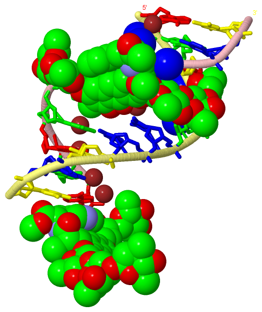
| 05 | AC5 | SOFTWARE | DG A:4 , DG A:5 , DC A:7 , ZN A:102 , HOH A:201 , DG B:5 , DC B:6 , DC B:7 , DT B:8 , DC B:9 , HOH B:202 , HOH B:205 , HOH B:206 , HOH B:212 , HOH B:216 , HOH B:222 , HOH B:227 , HOH B:228 , HOH B:230 , HOH B:242 , HOH B:254 , 6O6 C:101 , HOH C:208 | binding site for residue 6O6 B 101 |
| 06 | AC6 | SOFTWARE | DT A:10 , DA B:1 , HOH B:201 , HOH B:208 , HOH B:211 , HOH B:217 , HOH B:221 , HOH B:224 , HOH B:235 , HOH B:237 , HOH B:238 , HOH B:244 , DG C:4 , DG C:5 , 6O6 C:102 , ZN C:105 , HOH C:215 , DA D:3 , DG D:5 , DC D:6 , DC D:7 , DT D:8 , DC D:9 , HOH D:201 | binding site for residue 6O6 B 102 |
| 07 | AC7 | SOFTWARE | DG B:4 , ZN B:105 , HOH B:232 , HOH B:243 , HOH B:248 , HOH B:252 | binding site for residue ZN B 103 |
| 08 | AC8 | SOFTWARE | DA B:1 , HOH B:219 , HOH B:223 , HOH B:246 , HOH B:261 , HOH D:212 | binding site for residue ZN B 104 |
| 09 | AC9 | SOFTWARE | DG B:5 , ZN B:103 , HOH B:248 | binding site for residue ZN B 105 |
| 10 | AD1 | SOFTWARE | DG B:5 , HOH B:258 | binding site for residue NA B 106 |
| 11 | AD2 | SOFTWARE | DG A:5 , DC A:6 , DC A:7 , DT A:8 , ZN A:102 , HOH A:201 , DG B:4 , DG B:5 , 6O6 B:101 , DA C:1 , HOH C:201 , HOH C:202 , HOH C:208 , HOH C:210 , HOH C:214 , HOH C:228 , HOH C:235 , DT D:10 | binding site for residue 6O6 C 101 |
| 12 | AD3 | SOFTWARE | 6O6 B:102 , HOH B:201 , DG C:5 , DC C:6 , DC C:7 , DT C:8 , DC C:9 , ZN C:105 , NA C:109 , HOH C:207 , HOH C:213 , HOH C:215 , HOH C:217 , HOH C:218 , HOH C:229 , HOH C:234 , HOH C:236 , DG D:4 , DG D:5 , NA D:103 | binding site for residue 6O6 C 102 |
| 13 | AD4 | SOFTWARE | DG C:4 , ZN C:106 | binding site for residue ZN C 103 |
| 14 | AD5 | SOFTWARE | DA C:1 , ZN C:107 , HOH C:219 , HOH C:230 , HOH C:232 , HOH C:237 , HOH C:243 | binding site for residue ZN C 104 |
| 15 | AD6 | SOFTWARE | 6O6 B:102 , HOH B:201 , 6O6 C:102 , HOH C:215 | binding site for residue ZN C 105 |
| 16 | AD7 | SOFTWARE | DA C:3 , ZN C:103 | binding site for residue ZN C 106 |
| 17 | AD8 | SOFTWARE | DA C:1 , DG C:2 , ZN C:104 , HOH C:230 , HOH C:237 | binding site for residue ZN C 107 |
| 18 | AD9 | SOFTWARE | DC C:7 | binding site for residue NA C 108 |
| 19 | AE1 | SOFTWARE | DT C:8 , 6O6 C:102 , HOH C:218 | binding site for residue NA C 109 |
| 20 | AE2 | SOFTWARE | DG D:4 , HOH D:215 , HOH D:225 , HOH D:233 | binding site for residue ZN D 101 |
| 21 | AE3 | SOFTWARE | DA D:1 , HOH D:213 , HOH D:220 , HOH D:223 , HOH D:232 | binding site for residue ZN D 102 |
| 22 | AE4 | SOFTWARE | 6O6 C:102 , HOH C:213 , DA D:3 , HOH D:231 | binding site for residue NA D 103 |
| 23 | AE5 | SOFTWARE | DG D:5 | binding site for residue NA D 104 |
| 24 | AE6 | SOFTWARE | DA D:1 , DG D:2 , HOH D:226 , HOH D:236 | binding site for residue NA D 105 |
|
 Description