Show PDB file:   
         Plain Text   HTML   (compressed file size)
QuickSearch:   
by PDB,NDB,UniProt,PROSITE Code or Search Term(s)  
(-)Asym./Biol. Unit
(-)Asym./Biol. Unit - sites
collapse expand < >
Image Asym./Biol. Unit
Asym./Biol. Unit  (Jmol Viewer)
Image Asym./Biol. Unit - sites
Asym./Biol. Unit - sites  (Jmol Viewer)

(-) Description

Title :  CRYSTAL STRUCTURE OF THE MID, CAP-BINDING, MID-LINK AND 627 DOMAINS FROM AVIAN INFLUENZA A VIRUS POLYMERASE PB2 SUBUNIT BOUND TO M7GTP H32 CRYSTAL FORM
 
Authors :  E. Thierry, A. Pflug, D. Hart, S. Cusack
Date :  07 Nov 15  (Deposition) - 13 Jan 16  (Release) - 20 Jan 16  (Revision)
Method :  X-RAY DIFFRACTION
Resolution :  3.10
Chains :  Asym./Biol. Unit :  A
Keywords :  Viral Protein, Influenza A Pb2-C Region Containing Mid, Cap-Binding, Mid- Link, 627 And Nls Domains (Keyword Search: [Gene Ontology, PubMed, Web (Google))
 
Reference :  E. Thierry, D. Guilligay, J. Kosinski, T. Bock, S. Gaudon, A. Round, A. Pflug, N. Hengrung, K. El Omari, F. Baudin, D. J. Hart, M. Beck, S. Cusack
Influenza Polymerase Can Adopt An Alternative Configuration Involving A Radical Repacking Of Pb2 Domains.
Mol. Cell V. 61 125 2016
PubMed-ID: 26711008  |  Reference-DOI: 10.1016/J.MOLCEL.2015.11.016

(-) Compounds

Molecule 1 - INFLUENZA A PB2 SUBUNIT
    ChainsA
    EngineeredYES
    Expression SystemESCHERICHIA COLI
    Expression System StrainBL21
    Expression System Taxid511693
    Expression System VariantRIL
    Expression System VectorPET9
    Expression System Vector TypePLASMID
    FragmentPB2-C, RESIDUES 247-736
    Organism ScientificINFLUENZA A VIRUS
    Organism Taxid11320
    StrainA/VIETNAM/1203/2004(H5N1)

 Structural Features

(-) Chains, Units

  1
Asymmetric/Biological Unit A

Summary Information (see also Sequences/Alignments below)

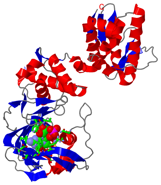
(-) Ligands, Modified Residues, Ions  (1, 1)

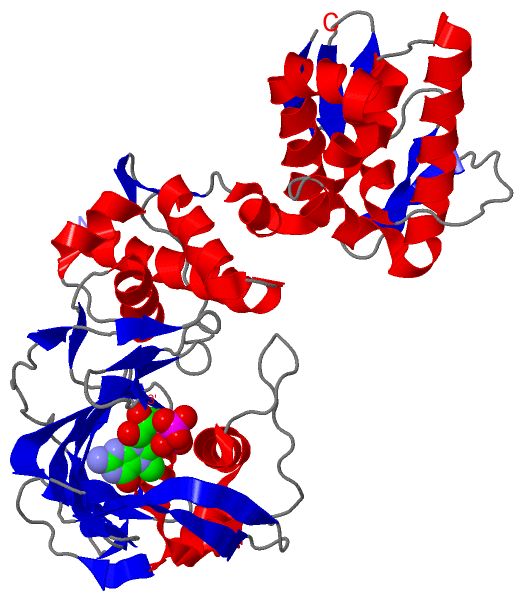
Asymmetric/Biological Unit (1, 1)
No.NameCountTypeFull Name
1MGT1Ligand/Ion7N-METHYL-8-HYDROGUANOSINE-5'-TRIPHOSPHATE

(-) Sites  (1, 1)

Asymmetric Unit (1, 1)
No.NameEvidenceResiduesDescription
1AC1SOFTWAREPHE A:323 , LYS A:339 , ARG A:355 , HIS A:357 , GLU A:361 , PHE A:363 , LYS A:376 , PHE A:404 , GLN A:406 , ASN A:429 , MET A:431 , ASN A:510 , VAL A:511BINDING SITE FOR RESIDUE MGT A1676

(-) SS Bonds  (0, 0)

(no "SS Bond" information available for 5FMQ)

(-) Cis Peptide Bonds  (0, 0)

(no "Cis Peptide Bond" information available for 5FMQ)

 Sequence-Structure Mapping

(-) SAPs(SNPs)/Variants  (0, 0)

(no "SAP(SNP)/Variant" information available for 5FMQ)

(-) PROSITE Motifs  (0, 0)

(no "PROSITE Motif" information available for 5FMQ)

(-) Exons   (0, 0)

(no "Exon" information available for 5FMQ)

(-) Sequences/Alignments

Asymmetric/Biological Unit
   Reformat: Number of residues per line =  ('0' or empty: single-line sequence representation)
  Number of residues per labelling interval =   
  UniProt sequence: complete  aligned part    
   Show mapping: SCOP domains CATH domains Pfam domains Secondary structure (by author)
SAPs(SNPs) PROSITE motifs Exons
(details for a mapped element are shown in a popup box when the mouse pointer rests over it)
Chain A from PDB  Type:PROTEIN  Length:424
                                                                                                                                                                                                                                                                                                                                                                                                                                                                        
               SCOP domains ---------------------------------------------------------------------------------------------------------------------------------------------------------------------------------------------------------------------------------------------------------------------------------------------------------------------------------------------------------------------------------------------------------------------------------------- SCOP domains
               CATH domains ---------------------------------------------------------------------------------------------------------------------------------------------------------------------------------------------------------------------------------------------------------------------------------------------------------------------------------------------------------------------------------------------------------------------------------------- CATH domains
               Pfam domains ---------------------------------------------------------------------------------------------------------------------------------------------------------------------------------------------------------------------------------------------------------------------------------------------------------------------------------------------------------------------------------------------------------------------------------------- Pfam domains
         Sec.struct. author hhhhhhhhhhhhhhhhhhhhhh.hhhhhhhhhhhhhee..eehhhhhhhh...hhhhhhhhhhh...ee..eee..eeeeeeee..eeeeeeee.....eeeeeeee..eeeeee...eeeeeeee..eeeeeeeee.hhhhhhhhhhhhhhhhhhhhhhhh...............hhhhhhhhhhhhhhhhhhhhhheee......eeee.....ee..eee..eeee...........ee..ee.....ee.....eee...............ee..hhhhhhhhhhhhhhhhhhhhhhhhhhhhhhhhhhhhhhhhh...hhhhhhhhhhhhhhhhhhhhhhhhhhhhhhh....hhhhhhhhhhhhh..........eeeee....eeeee.......eeee....eeee..eeeee. Sec.struct. author
                 SAPs(SNPs) ---------------------------------------------------------------------------------------------------------------------------------------------------------------------------------------------------------------------------------------------------------------------------------------------------------------------------------------------------------------------------------------------------------------------------------------- SAPs(SNPs)
                    PROSITE ---------------------------------------------------------------------------------------------------------------------------------------------------------------------------------------------------------------------------------------------------------------------------------------------------------------------------------------------------------------------------------------------------------------------------------------- PROSITE
                 Transcript ---------------------------------------------------------------------------------------------------------------------------------------------------------------------------------------------------------------------------------------------------------------------------------------------------------------------------------------------------------------------------------------------------------------------------------------- Transcript
                 5fmq A 252 NDDVDQSLIIAARNIVRRATVSADPLASLLEMCHSTQIGGIRMVDILRQNPTEEQAVDICKAAMGLRISSSFSFGGFTFKRTSGSSVKKEEEVLTGNLQTLKIRVHEGYEEFTMVGRRATAILRKATRRLIQLIVSGRDQQSIAEAIIVAMVFSQEDCMIKAVRGDLNFVNRANQRLNPMHQLLRHFQKDAKVLFQNWGIEPIDNVMGMIGILPDMTPSTEMSLRGVRVSKMGVDEYSSTERVVVSIDRFLRVRDQRGNVLLSPEEVSETQGTEKLTITYSSSMMWEINGPESVLVNTYQWIIRNWETVKIQWSQDPTMLYNKMEFEPFQSLVPKAARGQYSGFVRTLFQQMRDVLGTFDTVQIIKLLPFAAAPPKQSRMQFSSLTVNVRGSGMRILVRGNSPVFNYNKATKRLTVLGKDAGAL 675
                                   261       271       281       291       301       311       321       331       341       351       361       371       381       391       401       411       421       431       441       451       461       471       481       491       501       511       521       531       541       551       561       571       581       591       601       611       621       631       641       651       661       671    

   Legend:   → Mismatch (orange background)
  - → Gap (green background, '-', border residues have a numbering label)
    → Modified Residue (blue background, lower-case, 'x' indicates undefined single-letter code, labelled with number + name)
  x → Chemical Group (purple background, 'x', labelled with number + name, e.g. ACE or NH2)
  extra numbering lines below/above indicate numbering irregularities and modified residue names etc., number ends below/above '|'

 Classification and Annotation

(-) SCOP Domains  (0, 0)

(no "SCOP Domain" information available for 5FMQ)

(-) CATH Domains  (0, 0)

(no "CATH Domain" information available for 5FMQ)

(-) Pfam Domains  (0, 0)

(no "Pfam Domain" information available for 5FMQ)

(-) Gene Ontology  (15, 15)

Asymmetric/Biological Unit(hide GO term definitions)

 Visualization

(-) Interactive Views

Asymmetric/Biological Unit
  Complete Structure
    Jena3D(integrated viewing of ligand, site, SAP, PROSITE, SCOP information)
    WebMol | AstexViewer[tm]@PDBe
(Java Applets, require no local installation except for Java; loading may be slow)
    STRAP
(Java WebStart application, automatic local installation, requires Java; full application with system access!)
    RasMol
(require local installation)
    Molscript (VRML)
(requires installation of a VRML viewer; select preferred view via VRML and generate a mono or stereo PDF format file)
 
  Ligands, Modified Residues, Ions
    MGT  [ RasMol | Jena3D ]  +environment [ RasMol | Jena3D ]
 
  Sites
    AC1  [ RasMol ]  +environment [ RasMol ]
 
  Cis Peptide Bonds
(no "Cis Peptide Bonds" information available for 5fmq)
 

(-) Still Images

Jmol
  protein: cartoon or spacefill or dots and stick; nucleic acid: cartoon and stick; ligands: spacefill; active site: stick
Molscript
  protein, nucleic acid: cartoon; ligands: spacefill; active site: ball and stick

 Databases and Analysis Tools

(-) Databases

Access by PDB/NDB ID
  5fmq
    Family and Domain InformationProDom | SYSTERS
    General Structural InformationGlycoscienceDB | MMDB | NDB | OCA | PDB | PDBe | PDBj | PDBsum | PDBWiki | PQS | PROTEOPEDIA
    Orientation in MembranesOPM
    Protein SurfaceSURFACE
    Secondary StructureDSSP (structure derived) | HSSP (homology derived)
    Structural GenomicsGeneCensus
    Structural NeighboursCE | VAST
    Structure ClassificationCATH | Dali | SCOP
    Validation and Original DataBMRB Data View | BMRB Restraints Grid | EDS | PROCHECK | RECOORD | WHAT_CHECK
 
Access by UniProt ID/Accession number
  Q6DNN3_9INFA | Q6DNN3
    Comparative Protein Structure ModelsModBase
    Genomic InformationEnsembl
    Protein-protein InteractionDIP
    Sequence, Family and Domain InformationInterPro | Pfam | SMART | UniProtKB/TrEMBL
 
Access by Enzyme Classificator   (EC Number)
  (no 'Enzyme Classificator' available)
    General Enzyme InformationBRENDA | EC-PDB | Enzyme | IntEnz
    PathwayKEGG | MetaCyc
 
Access by Disease Identifier   (MIM ID)
  (no 'MIM ID' available)
    Disease InformationOMIM
 
Access by GenAge ID
  (no 'GenAge ID' available)
    Age Related InformationGenAge

(-) Analysis Tools

Access by PDB/NDB ID
    Domain InformationXDom
    Interatomic Contacts of Structural UnitsCSU
    Ligand-protein ContactsLPC
    Protein CavitiescastP
    Sequence and Secondary StructurePDBCartoon
    Structure AlignmentSTRAP(Java WebStart application, automatic local installation, requires Java; full application with system access!)
    Structure and Sequence BrowserSTING
 
Access by UniProt ID/Accession number
  Q6DNN3_9INFA | Q6DNN3
    Protein Disorder PredictionDisEMBL | FoldIndex | GLOBPLOT (for more information see DisProt)

 Related Entries

(-) Entries Sharing at Least One Protein Chain (UniProt ID)

UniProtKB/TrEMBL
        Q6DNN3_9INFA | Q6DNN33kc6 3l56 5fmm

(-) Related Entries Specified in the PDB File

5fml INFLUENZA POLYMERASE CAN ADOPT AN ALTERNATIVE CONFIGURATION INVOLVING A RADICAL REPACKING OF PB2 DOMAINS
5fmm INFLUENZA POLYMERASE CAN ADOPT AN ALTERNATIVE CONFIGURATION INVOLVING A RADICAL REPACKING OF PB2 DOMAINS