|
|
|
|
 Description
|
|
Compounds
|
||||||||||||||||||||||||||||||||||||||||||||
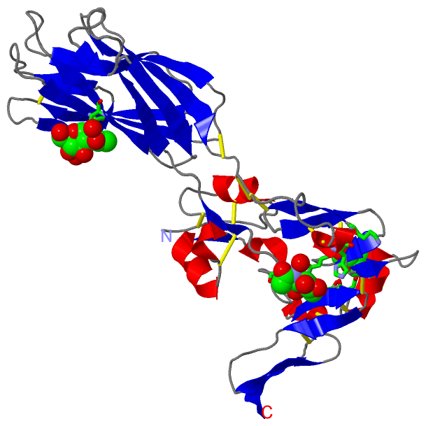
Chains, Units
Summary Information (see also Sequences/Alignments below) |
Ligands, Modified Residues, Ions (1, 3)
Asymmetric/Biological Unit (1, 3)
|
Sites (3, 3)
Asymmetric Unit (3, 3)
|
SS Bonds (13, 13)
Asymmetric/Biological Unit
|
||||||||||||||||||||||||||||||||||||||||||||||||||||||||
Cis Peptide Bonds (1, 1)
Asymmetric/Biological Unit
|
||||||||
SAPs(SNPs)/Variants (0, 0)| (no "SAP(SNP)/Variant" information available for 5E6X) |
PROSITE Motifs (0, 0)| (no "PROSITE Motif" information available for 5E6X) |
Exons (0, 0)| (no "Exon" information available for 5E6X) |
Sequences/Alignments
Asymmetric/Biological Unit
Chain A from PDB Type:PROTEIN Length:280
SCOP domains ---------------------------------------------------------------------------------------------------------------------------------------------------------------------------------------------------------------------------------------------------------------------------------------- SCOP domains
CATH domains ---------------------------------------------------------------------------------------------------------------------------------------------------------------------------------------------------------------------------------------------------------------------------------------- CATH domains
Pfam domains ---------------------------------------------------------------------------------------------------------------------------------------------------------------------------------------------------------------------------------------------------------------------------------------- Pfam domains
SAPs(SNPs) ---------------------------------------------------------------------------------------------------------------------------------------------------------------------------------------------------------------------------------------------------------------------------------------- SAPs(SNPs)
PROSITE ---------------------------------------------------------------------------------------------------------------------------------------------------------------------------------------------------------------------------------------------------------------------------------------- PROSITE
Transcript ---------------------------------------------------------------------------------------------------------------------------------------------------------------------------------------------------------------------------------------------------------------------------------------- Transcript
5e6x A 1 QECTKFKVSSCRECIESGPGCTWCQKLNFTGPGDPDSIRCDTRPQLLMRGCAADDIMDPTSLAETQEDHNGGQKQLSPQKVTLYLRPGQAAAFNVTFRRAKLSSRVFLDHNALPDTLKVTYDSFCSNGVTHRNQPRGDCDGVQINVPITFQVKVTATECIQEQSFVIRALGFTDIVTVQVLPQCECRCRDQSRDRSLCHGKGFLECGICRCDTGYIGKNCECQTQGRSSQELEGSCRKDNNSIICSGLGDCVCGQCLCHTSDVPGKLIYGQYCEHHHHHH 519
10 20 30 40 50 60 70 80 90 100| 349 359 369 379 389 399 409 419 429 439 449 459 469 479 489 499 509 519
100|
340
|
||||||||||||||||||||
SCOP Domains (0, 0)| (no "SCOP Domain" information available for 5E6X) |
CATH Domains (0, 0)| (no "CATH Domain" information available for 5E6X) |
Pfam Domains (0, 0)| (no "Pfam Domain" information available for 5E6X) |
Gene Ontology (46, 46)|
Asymmetric/Biological Unit(hide GO term definitions) |
Interactive Views
|
|||||||||||||||||||||||||||||||||||||||||||||||||||||||||||||||||||||||||||||||||||||||||||||||||||||||||||||||||||||||||||||||||||||
Still Images
|
||||||||||||||||
Databases
|
||||||||||||||||||||||||||||||||||||||||||||||||||||||||||||||||||||||||||||||||||||||||||||||||||||||||||||||||||||||||||||||||||||||||||||||||||||||||||||||||
Analysis Tools
|
|||||||||||||||||||||||||||||||||||||||||||||||||||||||||||||
Entries Sharing at Least One Protein Chain (UniProt ID)
Related Entries Specified in the PDB File
|
|