|
|
|
|
 Description
|
|
Compounds
|
||||||||||||||||||||||||||||||||||||||||||||
Chains, Units
Summary Information (see also Sequences/Alignments below) |
Ligands, Modified Residues, Ions (0, 0)| (no "Ligand,Modified Residues,Ions" information available for 5B1N) |
Sites (0, 0)| (no "Site" information available for 5B1N) |
SS Bonds (0, 0)| (no "SS Bond" information available for 5B1N) |
Cis Peptide Bonds (0, 0)| (no "Cis Peptide Bond" information available for 5B1N) |
SAPs(SNPs)/Variants (0, 0)| (no "SAP(SNP)/Variant" information available for 5B1N) |
PROSITE Motifs (0, 0)| (no "PROSITE Motif" information available for 5B1N) |
Exons (0, 0)| (no "Exon" information available for 5B1N) |
Sequences/Alignments
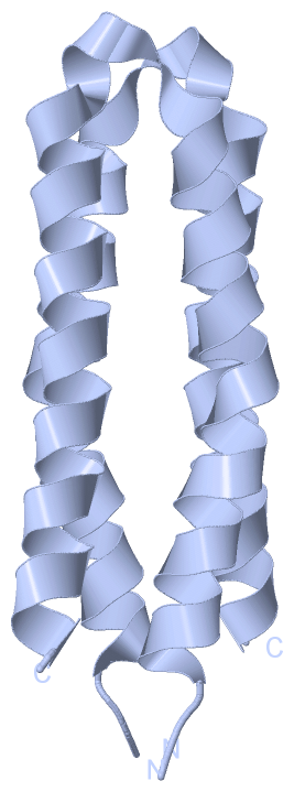
Asymmetric Unit
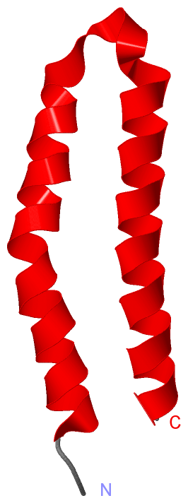
Chain A from PDB Type:PROTEIN Length:59
SCOP domains ----------------------------------------------------------- SCOP domains
CATH domains ----------------------------------------------------------- CATH domains
Pfam domains ----------------------------------------------------------- Pfam domains
SAPs(SNPs) ----------------------------------------------------------- SAPs(SNPs)
PROSITE ----------------------------------------------------------- PROSITE
Transcript ----------------------------------------------------------- Transcript
5b1n A 231 ADDRTLLMAGVSHDLRTPLTRIRLATEMMSEQDGYLAESINKDIEECNAIIEQFIDYLR 289
240 250 260 270 280
|
||||||||||||||||||||
SCOP Domains (0, 0)| (no "SCOP Domain" information available for 5B1N) |
CATH Domains (0, 0)| (no "CATH Domain" information available for 5B1N) |
Pfam Domains (0, 0)| (no "Pfam Domain" information available for 5B1N) |
Gene Ontology (24, 24)|
Asymmetric Unit(hide GO term definitions) |
Interactive Views
|
||||||||||||||||||||||||||||||||||||||||||||||||||||||||||||||||||||||||||||||||||||||||||||||||||||||||||||||||||||||||||||||||||||||
Still Images
|
||||||||||||||||
Databases
|
||||||||||||||||||||||||||||||||||||||||||||||||||||||||||||||||||||||||||||||||||||||||||||||||||||||||||||||||||||||||||||||||||||||||||||||||||||||||||||||||||||||||||||||||
Analysis Tools
|
|||||||||||||||||||||||||||||||||||||||||||||||||||||||||||||
Entries Sharing at Least One Protein Chain (UniProt ID)
Related Entries Specified in the PDB File
|
|