Show PDB file:   
         Plain Text   HTML   (compressed file size)
QuickSearch:   
by PDB,NDB,UniProt,PROSITE Code or Search Term(s)  
(-)Asymmetric Unit
(-)Asym. Unit - sites
(-)Biological Unit 1
(-)Biol. Unit 1 - sites
collapse expand < >
Image Asymmetric Unit
Asymmetric Unit  (Jmol Viewer)
Image Asym. Unit - sites
Asym. Unit - sites  (Jmol Viewer)
Image Biological Unit 1
Biological Unit 1  (Jmol Viewer)
Image Biol. Unit 1 - sites
Biol. Unit 1 - sites  (Jmol Viewer)

(-) Description

Title :  CRYSTAL STRUCTURES REVEAL TRANSIENT PERK LUMINAL DOMAIN TETRAMERIZATION IN ER STRESS SIGNALING
 
Authors :  M. Carrara, F. Prischi, M. M. U. Ali
Date :  25 Mar 15  (Deposition) - 13 May 15  (Release) - 17 Jun 15  (Revision)
Method :  X-RAY DIFFRACTION
Resolution :  3.14
Chains :  Asym. Unit :  A,B
Biol. Unit 1:  A,B  (2x)
Keywords :  Perk, Upr, Er Stress, Tetramer, Upr Activation, Upr Sensor, Unfolded Protein, Proteostasis, Signaling Protein (Keyword Search: [Gene Ontology, PubMed, Web (Google))
 
Reference :  M. Carrara, F. Prischi, P. R. Nowak, M. M. Ali
Crystal Structures Reveal Transient Perk Luminal Domain Tetramerization In Endoplasmic Reticulum Stress Signaling.
Embo J. V. 34 1589 2015
PubMed-ID: 25925385  |  Reference-DOI: 10.15252/EMBJ.201489183

(-) Compounds

Molecule 1 - EUKARYOTIC TRANSLATION INITIATION FACTOR 2-ALPHA KINASE 3
    ChainsA, B
    EC Number2.7.11.1
    EngineeredYES
    Expression SystemESCHERICHIA COLI
    Expression System Taxid562
    FragmentUNP RESIDUES 104-403
    GeneEIF2AK3, PEK, PERK
    Organism CommonHUMAN
    Organism ScientificHOMO SAPIENS
    Organism Taxid9606
    SynonymPRKR-LIKE ENDOPLASMIC RETICULUM KINASE,PANCREATIC EIF2-ALPHA KINASE,HSPEK

 Structural Features

(-) Chains, Units

  12
Asymmetric Unit AB
Biological Unit 1 (2x)AB

Summary Information (see also Sequences/Alignments below)

(-) Ligands, Modified Residues, Ions  (1, 2)

Asymmetric Unit (1, 2)
No.NameCountTypeFull Name
1W2Ligand/IonTUNGSTEN ION
Biological Unit 1 (0, 0)
No.NameCountTypeFull Name
1W-1Ligand/IonTUNGSTEN ION

(-) Sites  (1, 1)

Asymmetric Unit (1, 1)
No.NameEvidenceResiduesDescription
1AC1SOFTWARELYS B:256binding site for residue W B 501

(-) SS Bonds  (1, 1)

Asymmetric Unit
No.Residues
1A:216 -A:221

(-) Cis Peptide Bonds  (9, 9)

Asymmetric Unit
No.Residues
1Asp A:159 -Gly A:160
2Phe A:335 -Cys A:336
3Cys A:336 -Thr A:337
4Tyr A:391 -Arg A:392
5Arg A:392 -Gly A:393
6Glu B:121 -Asn B:122
7Ala B:207 -Tyr B:208
8Gly B:210 -Lys B:211
9Pro B:250 -Arg B:251

 Sequence-Structure Mapping

(-) SAPs(SNPs)/Variants  (0, 0)

(no "SAP(SNP)/Variant" information available for 4YZS)

(-) PROSITE Motifs  (0, 0)

(no "PROSITE Motif" information available for 4YZS)

(-) Exons   (0, 0)

(no "Exon" information available for 4YZS)

(-) Sequences/Alignments

Asymmetric Unit
   Reformat: Number of residues per line =  ('0' or empty: single-line sequence representation)
  Number of residues per labelling interval =   
  UniProt sequence: complete  aligned part    
   Show mapping: SCOP domains CATH domains Pfam domains Secondary structure (by author)
SAPs(SNPs) PROSITE motifs Exons
(details for a mapped element are shown in a popup box when the mouse pointer rests over it)
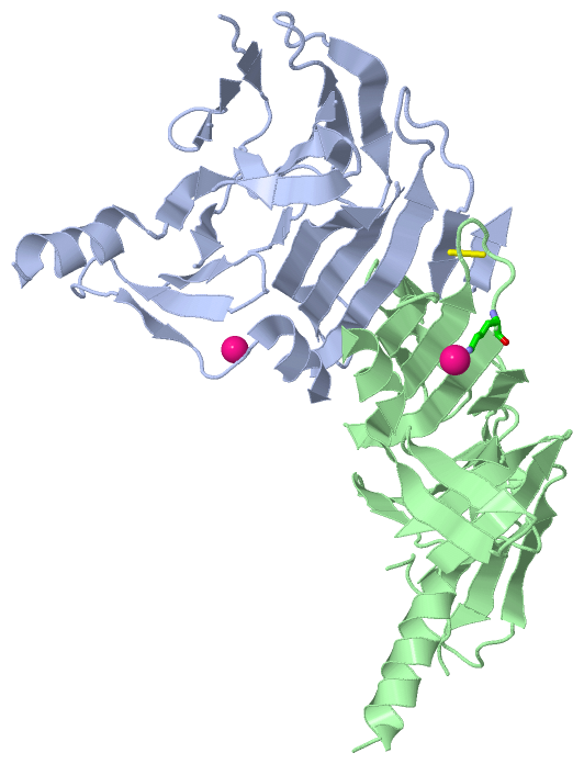
Chain A from PDB  Type:PROTEIN  Length:200
                                                                                                                                                                                                                                        
               SCOP domains -------------------------------------------------------------------------------------------------------------------------------------------------------------------------------------------------------- SCOP domains
               CATH domains -------------------------------------------------------------------------------------------------------------------------------------------------------------------------------------------------------- CATH domains
               Pfam domains -------------------------------------------------------------------------------------------------------------------------------------------------------------------------------------------------------- Pfam domains
         Sec.struct. author .eeeeee...eeeee.......eeeeee......ee....eeee.....eeee......eee...hhhhhhh..eeeeeeeeeeee..........eee..eee..eeeeeeeeeeee......eeeeeeeeeeeee..ee....ee....ee....eeeeeeee..eeee...hhhhhhhhhh..eeee....eeee.. Sec.struct. author
                 SAPs(SNPs) -------------------------------------------------------------------------------------------------------------------------------------------------------------------------------------------------------- SAPs(SNPs)
                    PROSITE -------------------------------------------------------------------------------------------------------------------------------------------------------------------------------------------------------- PROSITE
                 Transcript -------------------------------------------------------------------------------------------------------------------------------------------------------------------------------------------------------- Transcript
                 4yzs A 104 SLVIISTLDGRIAALDPENHGKKQWDLDVGSGSLVSSSLKMIIPSLDGALFQWDRDRESMETVPFTVESLLESVVLVGGKSLTTYGLSAYSGKVRYICSALGCRQLLLQRTQKTVRAVGPRSGNEKWNFSVGHFELRYKVSVADWKVMEWEYQFCTPIASAWLLKDGKVIPISIVEAARGATENSVYLGMYRGQLYLQSS 400
                                   113       123       133       152       162       172       182  ||   198       208       218    || 240       250       260       312       331       341       351  ||   380       390       400
                                                                142|                              185|                            223|                             268|      320|                     354|                          
                                                                 152                               192                             236                              311       330                      374                          

Chain B from PDB  Type:PROTEIN  Length:234
                                                                                                                                                                                                                                                                          
               SCOP domains ------------------------------------------------------------------------------------------------------------------------------------------------------------------------------------------------------------------------------------------ SCOP domains
               CATH domains ------------------------------------------------------------------------------------------------------------------------------------------------------------------------------------------------------------------------------------------ CATH domains
               Pfam domains ------------------------------------------------------------------------------------------------------------------------------------------------------------------------------------------------------------------------------------------ Pfam domains
         Sec.struct. author ..eeeee....eeeee.....ee...........eeee..eeee....eeeeee....eeeeeeeehhhhhh....eeeeeeeeeeeee......eeeee....eee....eeeeeeeeeeeee........eeeeeeeeeeee.......eeeeee....eeeeee.....eeeeee....eeeeeeee..eeee.........hhhhhhhhhhhhhheeeeee..eeeeee. Sec.struct. author
                 SAPs(SNPs) ------------------------------------------------------------------------------------------------------------------------------------------------------------------------------------------------------------------------------------------ SAPs(SNPs)
                    PROSITE ------------------------------------------------------------------------------------------------------------------------------------------------------------------------------------------------------------------------------------------ PROSITE
                 Transcript ------------------------------------------------------------------------------------------------------------------------------------------------------------------------------------------------------------------------------------------ Transcript
                 4yzs B 104 SLVIISTLDGRIAALDPENHGKKQWDLDVGSGSLVSSSLKMIIPSLDGALFQWDRDRESMETVPFTVESLLESDDVVLVGGKSLTTYGLSAYSGKVRYICSALGCRQWDSILLLQRTQKTVRAVGPRSGNEKWNFSVGHFELRYIPAAIMDIVIKVSVADWKVMAFSKKGGHLEWEYQFCTPIASAWLLKDGKVIPISLFDDTSEEDIVEAARGATENSVYLGMYRGQLYLQSS 400
                                   113       123       133       152       162       172       182  ||   196       206       216       226|      244       254       264     ||306       316       326       336       346       356   ||  376       386       396    
                                                                142|                              185|                                 226|                                270|                                                      360|                             
                                                                 152                               190                                  235                                 303                                                       371                             

   Legend:   → Mismatch (orange background)
  - → Gap (green background, '-', border residues have a numbering label)
    → Modified Residue (blue background, lower-case, 'x' indicates undefined single-letter code, labelled with number + name)
  x → Chemical Group (purple background, 'x', labelled with number + name, e.g. ACE or NH2)
  extra numbering lines below/above indicate numbering irregularities and modified residue names etc., number ends below/above '|'

 Classification and Annotation

(-) SCOP Domains  (0, 0)

(no "SCOP Domain" information available for 4YZS)

(-) CATH Domains  (0, 0)

(no "CATH Domain" information available for 4YZS)

(-) Pfam Domains  (0, 0)

(no "Pfam Domain" information available for 4YZS)

(-) Gene Ontology  (58, 58)

Asymmetric Unit(hide GO term definitions)

 Visualization

(-) Interactive Views

Asymmetric Unit
  Complete Structure
    Jena3D(integrated viewing of ligand, site, SAP, PROSITE, SCOP information)
    WebMol | AstexViewer[tm]@PDBe
(Java Applets, require no local installation except for Java; loading may be slow)
    STRAP
(Java WebStart application, automatic local installation, requires Java; full application with system access!)
    RasMol
(require local installation)
    Molscript (VRML)
(requires installation of a VRML viewer; select preferred view via VRML and generate a mono or stereo PDF format file)
 
  Ligands, Modified Residues, Ions
    W  [ RasMol | Jena3D ]  +environment [ RasMol | Jena3D ]
 
  Sites
    AC1  [ RasMol ]  +environment [ RasMol ]
 
  Cis Peptide Bonds
    Ala B:207 - Tyr B:208   [ RasMol ]  
    Arg A:392 - Gly A:393   [ RasMol ]  
    Asp A:159 - Gly A:160   [ RasMol ]  
    Cys A:336 - Thr A:337   [ RasMol ]  
    Glu B:121 - Asn B:122   [ RasMol ]  
    Gly B:210 - Lys B:211   [ RasMol ]  
    Phe A:335 - Cys A:336   [ RasMol ]  
    Pro B:250 - Arg B:251   [ RasMol ]  
    Tyr A:391 - Arg A:392   [ RasMol ]  
 
Biological Unit
  Complete Structure
    Biological Unit 1  [ Jena3D ]

(-) Still Images

Jmol
  protein: cartoon or spacefill or dots and stick; nucleic acid: cartoon and stick; ligands: spacefill; active site: stick
Molscript
  protein, nucleic acid: cartoon; ligands: spacefill; active site: ball and stick

 Databases and Analysis Tools

(-) Databases

Access by PDB/NDB ID
  4yzs
    Family and Domain InformationProDom | SYSTERS
    General Structural InformationGlycoscienceDB | MMDB | NDB | OCA | PDB | PDBe | PDBj | PDBsum | PDBWiki | PQS | PROTEOPEDIA
    Orientation in MembranesOPM
    Protein SurfaceSURFACE
    Secondary StructureDSSP (structure derived) | HSSP (homology derived)
    Structural GenomicsGeneCensus
    Structural NeighboursCE | VAST
    Structure ClassificationCATH | Dali | SCOP
    Validation and Original DataBMRB Data View | BMRB Restraints Grid | EDS | PROCHECK | RECOORD | WHAT_CHECK
 
Access by UniProt ID/Accession number
  E2AK3_HUMAN | Q9NZJ5
    Comparative Protein Structure ModelsModBase
    Genomic InformationEnsembl
    Protein-protein InteractionDIP
    Sequence, Family and Domain InformationInterPro | Pfam | SMART | UniProtKB/SwissProt
 
Access by Enzyme Classificator   (EC Number)
  2.7.11.1
    General Enzyme InformationBRENDA | EC-PDB | Enzyme | IntEnz
    PathwayKEGG | MetaCyc
 
Access by Disease Identifier   (MIM ID)
  (no 'MIM ID' available)
    Disease InformationOMIM
 
Access by GenAge ID
  (no 'GenAge ID' available)
    Age Related InformationGenAge

(-) Analysis Tools

Access by PDB/NDB ID
    Domain InformationXDom
    Interatomic Contacts of Structural UnitsCSU
    Ligand-protein ContactsLPC
    Protein CavitiescastP
    Sequence and Secondary StructurePDBCartoon
    Structure AlignmentSTRAP(Java WebStart application, automatic local installation, requires Java; full application with system access!)
    Structure and Sequence BrowserSTING
 
Access by UniProt ID/Accession number
  E2AK3_HUMAN | Q9NZJ5
    Protein Disorder PredictionDisEMBL | FoldIndex | GLOBPLOT (for more information see DisProt)

 Related Entries

(-) Entries Sharing at Least One Protein Chain (UniProt ID)

UniProtKB/Swiss-Prot
        E2AK3_HUMAN | Q9NZJ54g31 4g34 4m7i 4x7h 4x7j 4x7k 4x7l 4x7n 4x7o 5sv7

(-) Related Entries Specified in the PDB File

(no "Related Entries Specified in the PDB File" available for 4YZS)