|
|
|
|
 Description
|
|
Compounds
|
||||||||||||||||||||||||||||||||||||||||||||||||||||||||||||
Chains, Units
Summary Information (see also Sequences/Alignments below) |
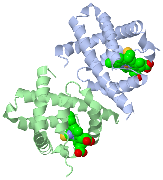
Ligands, Modified Residues, Ions (2, 4)| Asymmetric/Biological Unit (2, 4) |
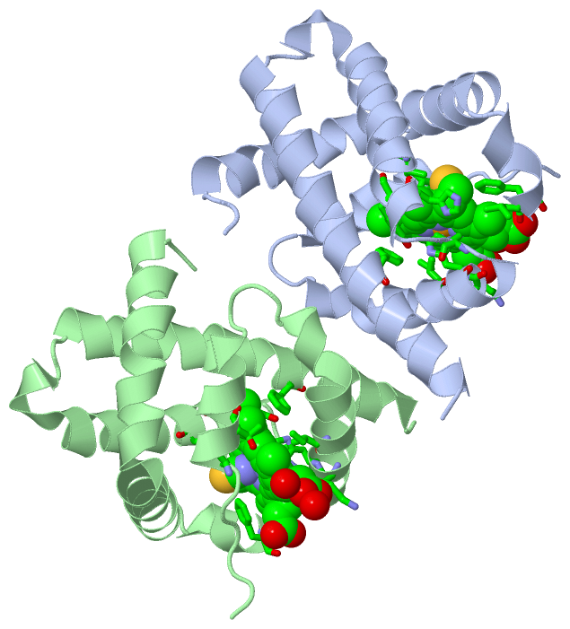
Sites (4, 4)
Asymmetric Unit (4, 4)
|
SS Bonds (0, 0)| (no "SS Bond" information available for 4VHB) |
Cis Peptide Bonds (0, 0)| (no "Cis Peptide Bond" information available for 4VHB) |
SAPs(SNPs)/Variants (0, 0)| (no "SAP(SNP)/Variant" information available for 4VHB) |
PROSITE Motifs (0, 0)| (no "PROSITE Motif" information available for 4VHB) |
Exons (0, 0)| (no "Exon" information available for 4VHB) |
Sequences/Alignments
Asymmetric/Biological Unit
Chain A from PDB Type:PROTEIN Length:138
SCOP domains d4vhba_ A: Bacterial dimeric hemoglobin SCOP domains
CATH domains 4vhbA00 A:1-146 Globins CATH domains
Pfam domains ------------------------------------------------------------------------------------------------------------------------------------------ Pfam domains
SAPs(SNPs) ------------------------------------------------------------------------------------------------------------------------------------------ SAPs(SNPs)
PROSITE ------------------------------------------------------------------------------------------------------------------------------------------ PROSITE
Transcript ------------------------------------------------------------------------------------------------------------------------------------------ Transcript
4vhb A 1 MLDQQTINIIKATVPVLKEHGVTITTTFYKNLFAKHPEVRPLFEQPKALAMTVLAAAQNIENLPAILPAVKKIAVKHCQAGVAAAHYPIVGQELLGAIKEVLGDAATDDILDAWGKAYGVIADVFIQVEADLYAQAVE 146
10 20 30 40 || 58 68 78 88 98 108 118 128 138
43|
52
Chain B from PDB Type:PROTEIN Length:137
SCOP domains d4vhbb_ B: Bacterial dimeric hemoglobin SCOP domains
CATH domains 4vhbB00 B:2-145 Globins CATH domains
Pfam domains ----------------------------------------------------------------------------------------------------------------------------------------- Pfam domains
SAPs(SNPs) ----------------------------------------------------------------------------------------------------------------------------------------- SAPs(SNPs)
PROSITE ----------------------------------------------------------------------------------------------------------------------------------------- PROSITE
Transcript ----------------------------------------------------------------------------------------------------------------------------------------- Transcript
4vhb B 2 LDQQTINIIKATVPVLKEHGVTITTTFYKNLFAKHPEVRPLFLEQPKALAMTVLAAAQNIENLPAILPAVKKIAVKHCQAGVAAAHYPIVGQELLGAIKEVLGDAATDDILDAWGKAYGVIADVFIQVEADLYAQAV 145
11 21 31 41 || 58 68 78 88 98 108 118 128 138
43|
51
|
||||||||||||||||||||
SCOP Domains (1, 2)
Asymmetric/Biological Unit
 |
CATH Domains (1, 2)
Asymmetric/Biological Unit
|
Pfam Domains (0, 0)| (no "Pfam Domain" information available for 4VHB) |
Gene Ontology (6, 6)|
Asymmetric/Biological Unit(hide GO term definitions) |
Interactive Views
|
||||||||||||||||||||||||||||||||||||||||||||||||||||||||||||||||||||||||||||||||||||||||||||||||||||||||||||||||||||||||||||||||||||||||||||||||||
Still Images
|
||||||||||||||||
Databases
|
||||||||||||||||||||||||||||||||||||||||||||||||||||||||||||||||||||||||||||||||||||||||||||||||||||||||||||||||||||||||||||||||||||||||||||||||||||||||||||||||
Analysis Tools
|
|||||||||||||||||||||||||||||||||||||||||||||||||||||||||||||
Entries Sharing at Least One Protein Chain (UniProt ID)
Related Entries Specified in the PDB File
|
|