|
|
|
|
 Description
|
|
Compounds
|
||||||||||||||||||||||||||||||||||||||||||||||||||||
Chains, Units
Summary Information (see also Sequences/Alignments below) |
Ligands, Modified Residues, Ions (2, 13)| Asymmetric Unit (2, 13) Biological Unit 1 (2, 26) |
Sites (11, 11)
Asymmetric Unit (11, 11)
|
SS Bonds (0, 0)| (no "SS Bond" information available for 3TM9) |
Cis Peptide Bonds (0, 0)| (no "Cis Peptide Bond" information available for 3TM9) |
SAPs(SNPs)/Variants (0, 0)| (no "SAP(SNP)/Variant" information available for 3TM9) |
PROSITE Motifs (1, 1)
Asymmetric Unit (1, 1)
|
||||||||||||||||||||||||||||||||||||||||||||||||
Exons (0, 0)| (no "Exon" information available for 3TM9) |
Sequences/Alignments
Asymmetric UnitChain A from PDB Type:PROTEIN Length:146 aligned with BAHG_VITST | P04252 from UniProtKB/Swiss-Prot Length:146 Alignment length:146 10 20 30 40 50 60 70 80 90 100 110 120 130 140 BAHG_VITST 1 MLDQQTINIIKATVPVLKEHGVTITTTFYKNLFAKHPEVRPLFDMGRQESLEQPKALAMTVLAAAQNIENLPAILPAVKKIAVKHCQAGVAAAHYPIVGQELLGAIKEVLGDAATDDILDAWGKAYGVIADVFIQVEADLYAQAVE 146 SCOP domains -------------------------------------------------------------------------------------------------------------------------------------------------- SCOP domains CATH domains -------------------------------------------------------------------------------------------------------------------------------------------------- CATH domains Pfam domains -------------------------------------------------------------------------------------------------------------------------------------------------- Pfam domains SAPs(SNPs) -------------------------------------------------------------------------------------------------------------------------------------------------- SAPs(SNPs) PROSITE GLOBIN PDB: A:1-135 UniProt: 1-135 ----------- PROSITE Transcript -------------------------------------------------------------------------------------------------------------------------------------------------- Transcript 3tm9 A 1 MLDQQTINIIKATVPVLKEHGVTITTTFAKNLFAKHPEVRPLFDMGRQESLEQPKALAMTVLAAAQNIENLPAILPAVKKIAVKHCQAGVAAAHYPIVGQELLGAIKEVLGDAATDDILDAWGKAYGVIADVFIQVEADLYAQAVE 146 10 20 30 40 50 60 70 80 90 100 110 120 130 140
|
||||||||||||||||||||
SCOP Domains (0, 0)| (no "SCOP Domain" information available for 3TM9) |
CATH Domains (0, 0)| (no "CATH Domain" information available for 3TM9) |
Pfam Domains (0, 0)| (no "Pfam Domain" information available for 3TM9) |
Gene Ontology (6, 6)|
Asymmetric Unit(hide GO term definitions) Chain A (BAHG_VITST | P04252)
|
||||||||||||||||||||||||||||||||||||||||||||||||
Interactive Views
|
|||||||||||||||||||||||||||||||||||||||||||||||||||||||||||||||||||||||||||||||||||||||||||||||||||||||||||||||||||||||||||||||||||||||||||||||||||||||||||||||||||||||||||||||||||||||||||||||||||||||||||||||||||||
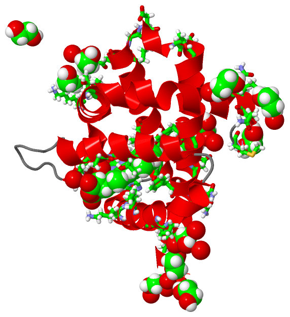
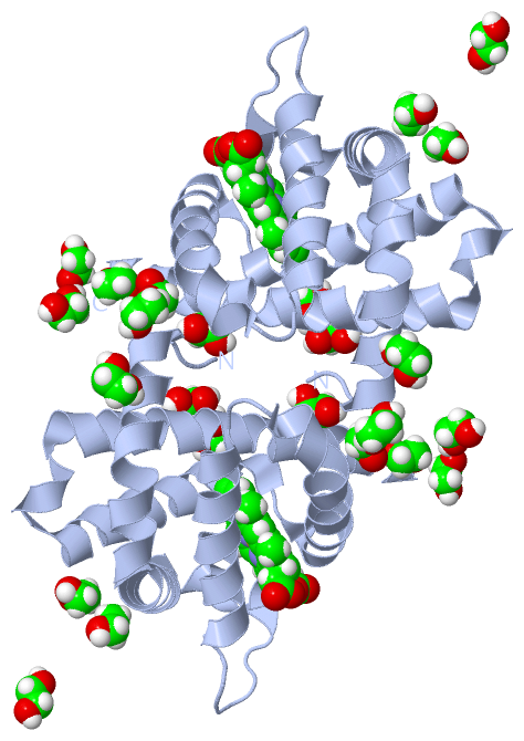
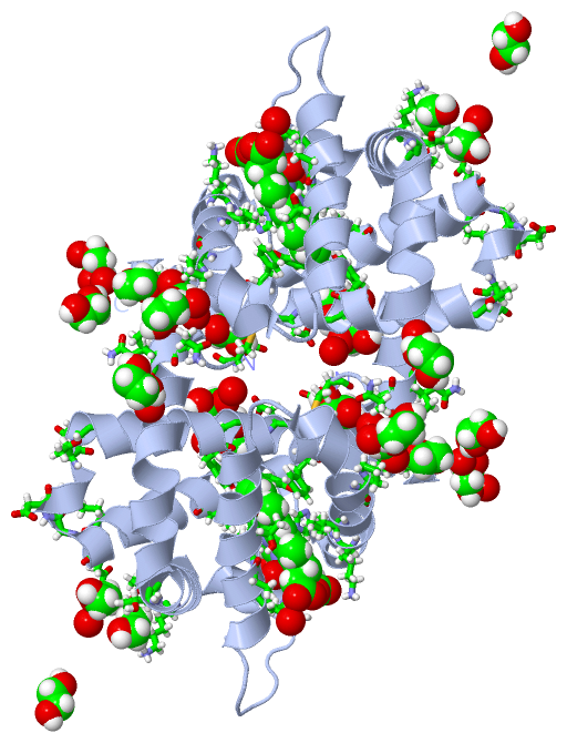
Still Images
|
||||||||||||||||
Databases
|
||||||||||||||||||||||||||||||||||||||||||||||||||||||||||||||||||||||||||||||||||||||||||||||||||||||||||||||||||||||||||||||||||||||||||||||||||||||||||||||||
Analysis Tools
|
|||||||||||||||||||||||||||||||||||||||||||||||||||||||||||||
Entries Sharing at Least One Protein Chain (UniProt ID)
Related Entries Specified in the PDB File
|
|