Show PDB file:   
         Plain Text   HTML   (compressed file size)
QuickSearch:   
by PDB,NDB,UniProt,PROSITE Code or Search Term(s)  
(-)Asym./Biol. Unit
(-)Asym./Biol. Unit - sites
collapse expand < >
Image Asym./Biol. Unit
Asym./Biol. Unit  (Jmol Viewer)
Image Asym./Biol. Unit - sites
Asym./Biol. Unit - sites  (Jmol Viewer)

(-) Description

Title :  THE STRUCTURE OF A-PGS FROM PSEUDOMONAS AERUGINOSA (SEMET DERIVATIVE)
 
Authors :  J. Krausze, S. Hebecker, T. Hasenkampf, D. W. Heinz, J. Moser
Date :  16 Oct 14  (Deposition) - 19 Aug 15  (Release) - 12 Jul 17  (Revision)
Method :  X-RAY DIFFRACTION
Resolution :  3.10
Chains :  Asym./Biol. Unit :  A
Keywords :  Transferase, Sad, Trna-Dependent Aminoacylation, Bacterial Resistance Proteins, A-Pgs, Lipid Homeostasis (Keyword Search: [Gene Ontology, PubMed, Web (Google))
 
Reference :  S. Hebecker, J. Krausze, T. Hasenkampf, J. Schneider, M. Groenewold, J. Reichelt, D. Jahn, D. W. Heinz, J. Moser
Structures Of Two Bacterial Resistance Factors Mediating Trna-Dependent Aminoacylation Of Phosphatidylglycerol With Lysine Or Alanine.
Proc. Natl. Acad. Sci. Usa V. 112 10691 2015
PubMed-ID: 26261323  |  Reference-DOI: 10.1073/PNAS.1511167112

(-) Compounds

Molecule 1 - ALANYL-TRNA-DEPENDENT L-ALANYL- PHOPHATIDYLGLYCEROL SYNTHASE
    AtccATCC 1569
    ChainsA
    EC Number2.3.2.11
    EngineeredYES
    Expression SystemESCHERICHIA COLI
    Expression System PlasmidPET52B
    Expression System StrainBL21(DE3)
    Expression System Taxid469008
    Expression System Vector TypePLASMID
    FragmentSOLUBLE DOMAIN, UNP RESIDUES 543-881
    MutationYES
    Organism ScientificPSEUDOMONAS AERUGINOSA
    Organism Taxid208964
    Other DetailsSELENO-METHIONINE DERIVATIVE
    StrainPAO1

 Structural Features

(-) Chains, Units

  1
Asymmetric/Biological Unit A

Summary Information (see also Sequences/Alignments below)

(-) Ligands, Modified Residues, Ions  (3, 14)

Asymmetric/Biological Unit (3, 14)
No.NameCountTypeFull Name
1CL3Ligand/IonCHLORIDE ION
2MSE9Mod. Amino AcidSELENOMETHIONINE
3SO42Ligand/IonSULFATE ION

(-) Sites  (5, 5)

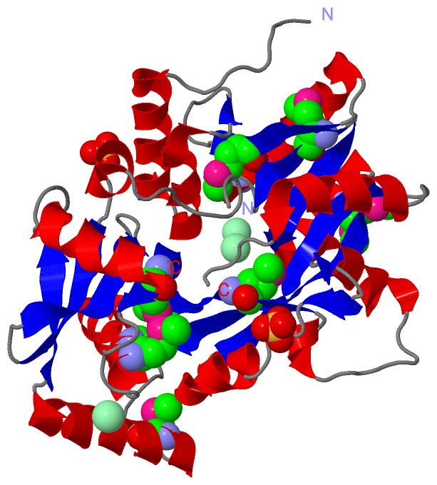
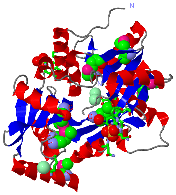
Asymmetric Unit (5, 5)
No.NameEvidenceResiduesDescription
1AC1SOFTWAREARG A:850BINDING SITE FOR RESIDUE CL A 1877
2AC2SOFTWAREGLU A:640BINDING SITE FOR RESIDUE CL A 1881
3AC3SOFTWARELYS A:775BINDING SITE FOR RESIDUE CL A 1878
4AC4SOFTWAREARG A:826 , TYR A:831 , ASN A:832 , PHE A:833 , GLN A:834BINDING SITE FOR RESIDUE SO4 A 1879
5AC5SOFTWAREARG A:562 , ARG A:565 , HIS A:566BINDING SITE FOR RESIDUE SO4 A 1880

(-) SS Bonds  (0, 0)

(no "SS Bond" information available for 4V34)

(-) Cis Peptide Bonds  (0, 0)

(no "Cis Peptide Bond" information available for 4V34)

 Sequence-Structure Mapping

(-) SAPs(SNPs)/Variants  (0, 0)

(no "SAP(SNP)/Variant" information available for 4V34)

(-) PROSITE Motifs  (0, 0)

(no "PROSITE Motif" information available for 4V34)

(-) Exons   (0, 0)

(no "Exon" information available for 4V34)

(-) Sequences/Alignments

Asymmetric/Biological Unit
   Reformat: Number of residues per line =  ('0' or empty: single-line sequence representation)
  Number of residues per labelling interval =   
  UniProt sequence: complete  aligned part    
   Show mapping: SCOP domains CATH domains Pfam domains Secondary structure (by author)
SAPs(SNPs) PROSITE motifs Exons
(details for a mapped element are shown in a popup box when the mouse pointer rests over it)
Chain A from PDB  Type:PROTEIN  Length:323
                                                                                                                                                                                                                                                                                                                                                                   
               SCOP domains ----------------------------------------------------------------------------------------------------------------------------------------------------------------------------------------------------------------------------------------------------------------------------------------------------------------------------------- SCOP domains
               CATH domains ----------------------------------------------------------------------------------------------------------------------------------------------------------------------------------------------------------------------------------------------------------------------------------------------------------------------------------- CATH domains
               Pfam domains ----------------------------------------------------------------------------------------------------------------------------------------------------------------------------------------------------------------------------------------------------------------------------------------------------------------------------------- Pfam domains
         Sec.struct. author .......hhhhhhhhhhhhhh..hhhhhhhhhh..eeee......eeeeeee..eeeee.....hhhhhhhhhhhhhhhhhhh..eeeeeeehhhhhhhhhhh.eeeeeeeeeeeee.........hhhhhhhhhhhhhhhhh.eeeeee.....hhhhhhhhhhhh........hhhhhh..eeeeeee..eeeeeeeee......eeeeeeeee......hhhhhhhhhhhhhhhhhh..eeeeeeee.hhh......hhhhhhhhhh.ee..ee....hhhhhhhhhh.eeeeeeeeee....hhhhhhhhhhhhh.... Sec.struct. author
                 SAPs(SNPs) ----------------------------------------------------------------------------------------------------------------------------------------------------------------------------------------------------------------------------------------------------------------------------------------------------------------------------------- SAPs(SNPs)
                    PROSITE ----------------------------------------------------------------------------------------------------------------------------------------------------------------------------------------------------------------------------------------------------------------------------------------------------------------------------------- PROSITE
                 Transcript ----------------------------------------------------------------------------------------------------------------------------------------------------------------------------------------------------------------------------------------------------------------------------------------------------------------------------------- Transcript
                 4v34 A 546 PPAIREPNAEELQRAARIIRHSDQPDGGLALTGDKALLFHESDDAFLmYARRGRSmIALYDPIGPAmQRAELIWQFRDLCDLHHARPVFYQVRAENLPFYmDIGLTALKLGEEARVDLLRFDLENAGAAmKDLRYTWNRGQRDGLALEFHEPGQAPLDELKAISDAWKGFSLGRFTPAYLNFFRIAIVRHQGKPVAFANLLETDSRELASLDLmRVHPDAPKLTmEFLmLGLILHYKAQGHARFSLGmVPLAGLQPRRGAPLTQRLGALVFRRGEQFYNFQGLRRFKDKFQPDWEPRYLAVPAGLDPLVALADTAALIAGGLT 876
                                   555       565       575       585       595     | 605      |615       625       635       645|      655       665       675       685       695       705      |723       733       743       753       763   |   773    |  783       793       803       813       823       833       843       853       863       873   
                                                                         593-MSE 601-MSE    612-MSE                           646-MSE                      675-MSE                              712|                                           767-MSE    778-MSE                801-MSE                                                                       
                                                                                                                                                                                                 721                                                          782-MSE                                                                                          

   Legend:   → Mismatch (orange background)
  - → Gap (green background, '-', border residues have a numbering label)
    → Modified Residue (blue background, lower-case, 'x' indicates undefined single-letter code, labelled with number + name)
  x → Chemical Group (purple background, 'x', labelled with number + name, e.g. ACE or NH2)
  extra numbering lines below/above indicate numbering irregularities and modified residue names etc., number ends below/above '|'

 Classification and Annotation

(-) SCOP Domains  (0, 0)

(no "SCOP Domain" information available for 4V34)

(-) CATH Domains  (0, 0)

(no "CATH Domain" information available for 4V34)

(-) Pfam Domains  (0, 0)

(no "Pfam Domain" information available for 4V34)

(-) Gene Ontology  (2, 2)

Asymmetric/Biological Unit(hide GO term definitions)

 Visualization

(-) Interactive Views

Asymmetric/Biological Unit
  Complete Structure
    Jena3D(integrated viewing of ligand, site, SAP, PROSITE, SCOP information)
    WebMol | AstexViewer[tm]@PDBe
(Java Applets, require no local installation except for Java; loading may be slow)
    STRAP
(Java WebStart application, automatic local installation, requires Java; full application with system access!)
    RasMol
(require local installation)
    Molscript (VRML)
(requires installation of a VRML viewer; select preferred view via VRML and generate a mono or stereo PDF format file)
 
  Ligands, Modified Residues, Ions
    CL  [ RasMol | Jena3D ]  +environment [ RasMol | Jena3D ]
    MSE  [ RasMol | Jena3D ]  +environment [ RasMol | Jena3D ]
    SO4  [ RasMol | Jena3D ]  +environment [ RasMol | Jena3D ]
 
  Sites
    AC1  [ RasMol ]  +environment [ RasMol ]
    AC2  [ RasMol ]  +environment [ RasMol ]
    AC3  [ RasMol ]  +environment [ RasMol ]
    AC4  [ RasMol ]  +environment [ RasMol ]
    AC5  [ RasMol ]  +environment [ RasMol ]
 
  Cis Peptide Bonds
(no "Cis Peptide Bonds" information available for 4v34)
 

(-) Still Images

Jmol
  protein: cartoon or spacefill or dots and stick; nucleic acid: cartoon and stick; ligands: spacefill; active site: stick
Molscript
  protein, nucleic acid: cartoon; ligands: spacefill; active site: ball and stick

 Databases and Analysis Tools

(-) Databases

Access by PDB/NDB ID
  4v34
    Family and Domain InformationProDom | SYSTERS
    General Structural InformationGlycoscienceDB | MMDB | NDB | OCA | PDB | PDBe | PDBj | PDBsum | PDBWiki | PQS | PROTEOPEDIA
    Orientation in MembranesOPM
    Protein SurfaceSURFACE
    Secondary StructureDSSP (structure derived) | HSSP (homology derived)
    Structural GenomicsGeneCensus
    Structural NeighboursCE | VAST
    Structure ClassificationCATH | Dali | SCOP
    Validation and Original DataBMRB Data View | BMRB Restraints Grid | EDS | PROCHECK | RECOORD | WHAT_CHECK
 
Access by UniProt ID/Accession number
  Q9I537_PSEAE | Q9I537
    Comparative Protein Structure ModelsModBase
    Genomic InformationEnsembl
    Protein-protein InteractionDIP
    Sequence, Family and Domain InformationInterPro | Pfam | SMART | UniProtKB/TrEMBL
 
Access by Enzyme Classificator   (EC Number)
  2.3.2.11
    General Enzyme InformationBRENDA | EC-PDB | Enzyme | IntEnz
    PathwayKEGG | MetaCyc
 
Access by Disease Identifier   (MIM ID)
  (no 'MIM ID' available)
    Disease InformationOMIM
 
Access by GenAge ID
  (no 'GenAge ID' available)
    Age Related InformationGenAge

(-) Analysis Tools

Access by PDB/NDB ID
    Domain InformationXDom
    Interatomic Contacts of Structural UnitsCSU
    Ligand-protein ContactsLPC
    Protein CavitiescastP
    Sequence and Secondary StructurePDBCartoon
    Structure AlignmentSTRAP(Java WebStart application, automatic local installation, requires Java; full application with system access!)
    Structure and Sequence BrowserSTING
 
Access by UniProt ID/Accession number
  Q9I537_PSEAE | Q9I537
    Protein Disorder PredictionDisEMBL | FoldIndex | GLOBPLOT (for more information see DisProt)

 Related Entries

(-) Entries Sharing at Least One Protein Chain (UniProt ID)

UniProtKB/TrEMBL
        Q9I537_PSEAE | Q9I5374v35

(-) Related Entries Specified in the PDB File

4v35 THE STRUCTURE OF A-PGS FROM PSEUDOMONAS AERUGINOSA
4v36 THE STRUCTURE OF L-PGS FROM BACILLUS LICHENIFORMIS