Show PDB file:   
         Plain Text   HTML   (compressed file size)
QuickSearch:   
by PDB,NDB,UniProt,PROSITE Code or Search Term(s)  
(-)Asym./Biol. Unit
(-)Asym./Biol. Unit - sites
collapse expand < >
Image Asym./Biol. Unit
Asym./Biol. Unit  (Jmol Viewer)
Image Asym./Biol. Unit - sites
Asym./Biol. Unit - sites  (Jmol Viewer)

(-) Description

Title :  CRYSTAL STRUCTURE OF HUMAN CAPERALPHA U2AF HOMOLOGY MOTIF (APO-STATE)
 
Authors :  S. Loerch, C. L. Kielkopf
Date :  12 Feb 14  (Deposition) - 14 May 14  (Release) - 01 Oct 14  (Revision)
Method :  X-RAY DIFFRACTION
Resolution :  2.20
Chains :  Asym./Biol. Unit :  A,B
Keywords :  U2Af Homology Motif, Uhm, Protein-Peptide Complex, Pre-Mrna Splicing Factor (Keyword Search: [Gene Ontology, PubMed, Web (Google))
 
Reference :  S. Loerch, A. Maucuer, V. Manceau, M. R. Green, C. L. Kielkopf
Cancer-Relevant Splicing Factor Caper Alpha Engages The Essential Splicing Factor Sf3B155 In A Specific Ternary Complex.
J. Biol. Chem. V. 289 17325 2014
PubMed-ID: 24795046  |  Reference-DOI: 10.1074/JBC.M114.558825

(-) Compounds

Molecule 1 - RNA-BINDING PROTEIN 39
    ChainsA, B
    EngineeredYES
    Expression SystemESCHERICHIA COLI
    Expression System PlasmidPGEX-6P
    Expression System StrainBL21 ROSETTA
    Expression System Taxid511693
    Expression System Vector TypePLASMID
    Fragment417-530
    GeneCAPER, CAPERALPHA, FSAP59, HCC1, RBM39, RNPC2
    Organism CommonHUMAN
    Organism ScientificHOMO SAPIENS
    Organism Taxid9606
    SynonymHEPATOCELLULAR CARCINOMA PROTEIN 1, RNA-BINDING MOTIF PROTEIN 39, RNA-BINDING REGION-CONTAINING PROTEIN 2, SPLICING FACTOR HCC1
    VariantISOFORM B

 Structural Features

(-) Chains, Units

  12
Asymmetric/Biological Unit AB

Summary Information (see also Sequences/Alignments below)

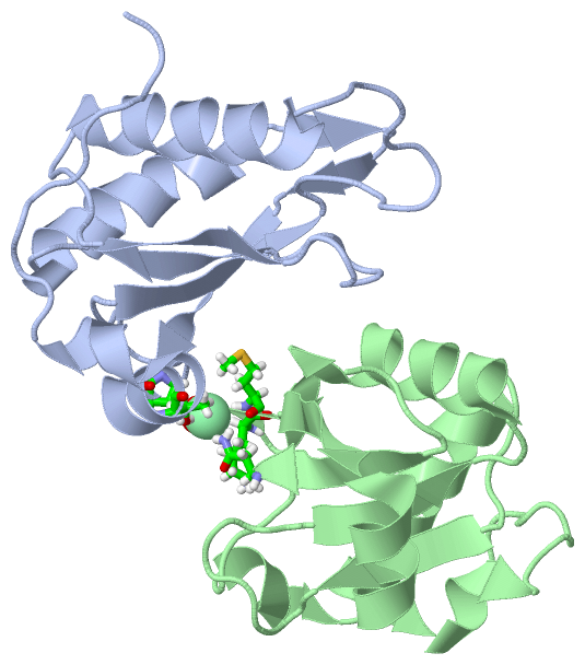
(-) Ligands, Modified Residues, Ions  (1, 1)

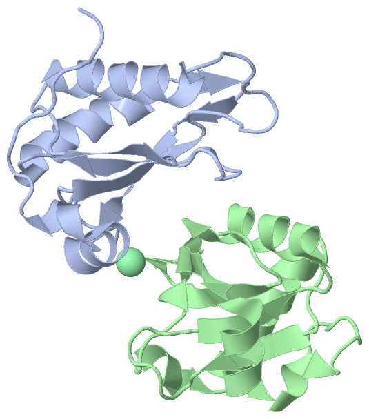
Asymmetric/Biological Unit (1, 1)
No.NameCountTypeFull Name
1CL1Ligand/IonCHLORIDE ION

(-) Sites  (1, 1)

Asymmetric Unit (1, 1)
No.NameEvidenceResiduesDescription
1AC1SOFTWAREPRO A:501 , THR A:504 , ASN B:424 , LYS B:493 , MET B:494 , HOH B:659binding site for residue CL A 601

(-) SS Bonds  (0, 0)

(no "SS Bond" information available for 4OZ0)

(-) Cis Peptide Bonds  (0, 0)

(no "Cis Peptide Bond" information available for 4OZ0)

 Sequence-Structure Mapping

(-) SAPs(SNPs)/Variants  (0, 0)

(no "SAP(SNP)/Variant" information available for 4OZ0)

(-) PROSITE Motifs  (0, 0)

(no "PROSITE Motif" information available for 4OZ0)

(-) Exons   (0, 0)

(no "Exon" information available for 4OZ0)

(-) Sequences/Alignments

Asymmetric/Biological Unit
   Reformat: Number of residues per line =  ('0' or empty: single-line sequence representation)
  Number of residues per labelling interval =   
  UniProt sequence: complete  aligned part    
   Show mapping: SCOP domains CATH domains Pfam domains Secondary structure (by author)
SAPs(SNPs) PROSITE motifs Exons
(details for a mapped element are shown in a popup box when the mouse pointer rests over it)
Chain A from PDB  Type:PROTEIN  Length:113
                                                                                                                                                 
               SCOP domains ----------------------------------------------------------------------------------------------------------------- SCOP domains
               CATH domains ----------------------------------------------------------------------------------------------------------------- CATH domains
               Pfam domains ----------------------------------------------------------------------------------------------------------------- Pfam domains
         Sec.struct. author .......eeeee...........hhhhhhhhhhhhhhh......eeee........eeee..hhhhhhhhhhhhh..ee..ee.eeeeehhhhhhhhhhhhh........... Sec.struct. author
                 SAPs(SNPs) ----------------------------------------------------------------------------------------------------------------- SAPs(SNPs)
                    PROSITE ----------------------------------------------------------------------------------------------------------------- PROSITE
                 Transcript ----------------------------------------------------------------------------------------------------------------- Transcript
                 4oz0 A 412 VQPLATQCFQLSNMFNPQTEEEVGWDTEIKDDVIEECNKHGGVIHIYVDKNSAQGNVYVKCPSIAAAIAAVNALHGRWFAGKMITAAYVPLPTYHNLFPDSMTATQLLVPSRR 524
                                   421       431       441       451       461       471       481       491       501       511       521   

Chain B from PDB  Type:PROTEIN  Length:108
                                                                                                                                            
               SCOP domains ------------------------------------------------------------------------------------------------------------ SCOP domains
               CATH domains ------------------------------------------------------------------------------------------------------------ CATH domains
               Pfam domains ------------------------------------------------------------------------------------------------------------ Pfam domains
         Sec.struct. author ....eeeee.....hhhhh.hhhhhhhhhhhhhhh......eeee........eeee..hhhhhhhhhhhhh..ee..ee.eeeeehhhhhhhhhhhhh......... Sec.struct. author
                 SAPs(SNPs) ------------------------------------------------------------------------------------------------------------ SAPs(SNPs)
                    PROSITE ------------------------------------------------------------------------------------------------------------ PROSITE
                 Transcript ------------------------------------------------------------------------------------------------------------ Transcript
                 4oz0 B 415 LATQCFQLSNMFNPQTEEEVGWDTEIKDDVIEECNKHGGVIHIYVDKNSAQGNVYVKCPSIAAAIAAVNALHGRWFAGKMITAAYVPLPTYHNLFPDSMTATQLLVPS 522
                                   424       434       444       454       464       474       484       494       504       514        

   Legend:   → Mismatch (orange background)
  - → Gap (green background, '-', border residues have a numbering label)
    → Modified Residue (blue background, lower-case, 'x' indicates undefined single-letter code, labelled with number + name)
  x → Chemical Group (purple background, 'x', labelled with number + name, e.g. ACE or NH2)
  extra numbering lines below/above indicate numbering irregularities and modified residue names etc., number ends below/above '|'

 Classification and Annotation

(-) SCOP Domains  (0, 0)

(no "SCOP Domain" information available for 4OZ0)

(-) CATH Domains  (0, 0)

(no "CATH Domain" information available for 4OZ0)

(-) Pfam Domains  (0, 0)

(no "Pfam Domain" information available for 4OZ0)

(-) Gene Ontology  (14, 14)

Asymmetric/Biological Unit(hide GO term definitions)

 Visualization

(-) Interactive Views

Asymmetric/Biological Unit
  Complete Structure
    Jena3D(integrated viewing of ligand, site, SAP, PROSITE, SCOP information)
    WebMol | AstexViewer[tm]@PDBe
(Java Applets, require no local installation except for Java; loading may be slow)
    STRAP
(Java WebStart application, automatic local installation, requires Java; full application with system access!)
    RasMol
(require local installation)
    Molscript (VRML)
(requires installation of a VRML viewer; select preferred view via VRML and generate a mono or stereo PDF format file)
 
  Ligands, Modified Residues, Ions
    CL  [ RasMol | Jena3D ]  +environment [ RasMol | Jena3D ]
 
  Sites
    AC1  [ RasMol ]  +environment [ RasMol ]
 
  Cis Peptide Bonds
(no "Cis Peptide Bonds" information available for 4oz0)
 

(-) Still Images

Jmol
  protein: cartoon or spacefill or dots and stick; nucleic acid: cartoon and stick; ligands: spacefill; active site: stick
Molscript
  protein, nucleic acid: cartoon; ligands: spacefill; active site: ball and stick

 Databases and Analysis Tools

(-) Databases

Access by PDB/NDB ID
  4oz0
    Family and Domain InformationProDom | SYSTERS
    General Structural InformationGlycoscienceDB | MMDB | NDB | OCA | PDB | PDBe | PDBj | PDBsum | PDBWiki | PQS | PROTEOPEDIA
    Orientation in MembranesOPM
    Protein SurfaceSURFACE
    Secondary StructureDSSP (structure derived) | HSSP (homology derived)
    Structural GenomicsGeneCensus
    Structural NeighboursCE | VAST
    Structure ClassificationCATH | Dali | SCOP
    Validation and Original DataBMRB Data View | BMRB Restraints Grid | EDS | PROCHECK | RECOORD | WHAT_CHECK
 
Access by UniProt ID/Accession number
  RBM39_HUMAN | Q14498
    Comparative Protein Structure ModelsModBase
    Genomic InformationEnsembl
    Protein-protein InteractionDIP
    Sequence, Family and Domain InformationInterPro | Pfam | SMART | UniProtKB/SwissProt
 
Access by Enzyme Classificator   (EC Number)
  (no 'Enzyme Classificator' available)
    General Enzyme InformationBRENDA | EC-PDB | Enzyme | IntEnz
    PathwayKEGG | MetaCyc
 
Access by Disease Identifier   (MIM ID)
  (no 'MIM ID' available)
    Disease InformationOMIM
 
Access by GenAge ID
  (no 'GenAge ID' available)
    Age Related InformationGenAge

(-) Analysis Tools

Access by PDB/NDB ID
    Domain InformationXDom
    Interatomic Contacts of Structural UnitsCSU
    Ligand-protein ContactsLPC
    Protein CavitiescastP
    Sequence and Secondary StructurePDBCartoon
    Structure AlignmentSTRAP(Java WebStart application, automatic local installation, requires Java; full application with system access!)
    Structure and Sequence BrowserSTING
 
Access by UniProt ID/Accession number
  RBM39_HUMAN | Q14498
    Protein Disorder PredictionDisEMBL | FoldIndex | GLOBPLOT (for more information see DisProt)

 Related Entries

(-) Entries Sharing at Least One Protein Chain (UniProt ID)

UniProtKB/Swiss-Prot
        RBM39_HUMAN | Q144982jrs 2mhn 4oo6 4oz1 4yud

(-) Related Entries Specified in the PDB File

(no "Related Entries Specified in the PDB File" available for 4OZ0)