Show PDB file:   
         Plain Text   HTML   (compressed file size)
QuickSearch:   
by PDB,NDB,UniProt,PROSITE Code or Search Term(s)  
(-)Asymmetric Unit
(-)Asym. Unit - sites
(-)Biological Unit 1
(-)Biol. Unit 1 - sites
(-)Biological Unit 2
(-)Biological Unit 3
collapse expand < >
Image Asymmetric Unit
Asymmetric Unit  (Jmol Viewer)
Image Asym. Unit - sites
Asym. Unit - sites  (Jmol Viewer)
Image Biological Unit 1
Biological Unit 1  (Jmol Viewer)
Image Biol. Unit 1 - sites
Biol. Unit 1 - sites  (Jmol Viewer)
Image Biological Unit 2
Biological Unit 2  (Jmol Viewer)
Image Biological Unit 3
Biological Unit 3  (Jmol Viewer)

(-) Description

Title :  M. LOTI CYCLIC-NUCLEOTIDE BINDING DOMAIN MUTANT DISPLAYING INVERTED LIGAND SELECTIVITY, CYCLIC-GMP BOUND
 
Authors :  F. Fonseca, J. Pessoa, J. H. Morais-Cabral
Date :  23 Sep 13  (Deposition) - 25 Jun 14  (Release) - 16 Jul 14  (Revision)
Method :  X-RAY DIFFRACTION
Resolution :  1.25
Chains :  Asym. Unit :  A,B
Biol. Unit 1:  A  (1x)
Biol. Unit 2:  B  (1x)
Biol. Unit 3:  A,B  (1x)
Keywords :  Mlotik1 Cnbd Mutant, Cgmp-Complex, Cyclic-Nucleotide Binding, Membrane Protein Domain, Metal Transport, Nucleotide Binding Protein (Keyword Search: [Gene Ontology, PubMed, Web (Google))
 
Reference :  J. Pessoa, F. Fonseca, S. Furini, J. H. Morais-Cabral
Determinants Of Ligand Selectivity In A Cyclic Nucleotide-Regulated Potassium Channel.
J. Gen. Physiol. V. 144 41 2014
PubMed-ID: 24981229  |  Reference-DOI: 10.1085/JGP.201311145

(-) Compounds

Molecule 1 - CYCLIC NUCLEOTIDE-GATED POTASSIUM CHANNEL MLL3241
    ChainsA, B
    EngineeredYES
    Expression SystemESCHERICHIA COLI
    Expression System PlasmidPGEX2T
    Expression System StrainBL21-CODONPLUS(DE3)-RP
    Expression System Taxid469008
    Expression System Vector TypePLASMID
    FragmentCYCLIC NUCLEOTIDE BINDING DOMAIN, UNP RESIDUES 216-355
    GeneMLL3241
    MutationYES
    Organism ScientificMESORHIZOBIUM LOTI
    Organism Taxid266835
    StrainMAFF303099
    SynonymMLOTIK1 CHANNEL

 Structural Features

(-) Chains, Units

  12
Asymmetric Unit AB
Biological Unit 1 (1x)A 
Biological Unit 2 (1x) B
Biological Unit 3 (1x)AB

Summary Information (see also Sequences/Alignments below)
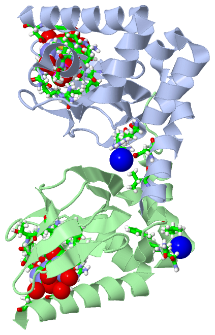
(-) Ligands, Modified Residues, Ions  (2, 4)

Asymmetric Unit (2, 4)
No.NameCountTypeFull Name
1NA2Ligand/IonSODIUM ION
2PCG2Ligand/IonCYCLIC GUANOSINE MONOPHOSPHATE
Biological Unit 1 (1, 1)
No.NameCountTypeFull Name
1NA-1Ligand/IonSODIUM ION
2PCG1Ligand/IonCYCLIC GUANOSINE MONOPHOSPHATE
Biological Unit 2 (1, 1)
No.NameCountTypeFull Name
1NA-1Ligand/IonSODIUM ION
2PCG1Ligand/IonCYCLIC GUANOSINE MONOPHOSPHATE
Biological Unit 3 (1, 2)
No.NameCountTypeFull Name
1NA-1Ligand/IonSODIUM ION
2PCG2Ligand/IonCYCLIC GUANOSINE MONOPHOSPHATE

(-) Sites  (4, 4)

Asymmetric Unit (4, 4)
No.NameEvidenceResiduesDescription
1AC1SOFTWARESER A:284 , PHE A:296 , GLY A:297 , GLU A:298 , MET A:299 , ALA A:300 , ARG A:307 , SER A:308 , ALA A:309 , VAL A:311 , ARG A:348 , ASP A:352 , HOH A:501 , HOH A:506 , HOH A:508 , HOH A:519 , HOH A:531BINDING SITE FOR RESIDUE PCG A 401
2AC2SOFTWAREVAL A:218 , ASP A:222 , ARG A:252 , ARG A:254 , HOH A:573 , HOH B:529BINDING SITE FOR RESIDUE NA A 402
3AC3SOFTWAREPHE B:296 , GLY B:297 , GLU B:298 , MET B:299 , ALA B:300 , ARG B:307 , SER B:308 , ALA B:309 , VAL B:311 , ARG B:348 , ASP B:352 , HOH B:501 , HOH B:506 , HOH B:507 , HOH B:509 , HOH B:512BINDING SITE FOR RESIDUE PCG B 401
4AC4SOFTWAREPHE B:236 , GLN B:237 , LEU B:239 , LEU B:244 , THR B:310BINDING SITE FOR RESIDUE NA B 402

(-) SS Bonds  (0, 0)

(no "SS Bond" information available for 4MUV)

(-) Cis Peptide Bonds  (0, 0)

(no "Cis Peptide Bond" information available for 4MUV)

 Sequence-Structure Mapping

(-) SAPs(SNPs)/Variants  (0, 0)

(no "SAP(SNP)/Variant" information available for 4MUV)

(-) PROSITE Motifs  (0, 0)

(no "PROSITE Motif" information available for 4MUV)

(-) Exons   (0, 0)

(no "Exon" information available for 4MUV)

(-) Sequences/Alignments

Asymmetric Unit
   Reformat: Number of residues per line =  ('0' or empty: single-line sequence representation)
  Number of residues per labelling interval =   
  UniProt sequence: complete  aligned part    
   Show mapping: SCOP domains CATH domains Pfam domains Secondary structure (by author)
SAPs(SNPs) PROSITE motifs Exons
(details for a mapped element are shown in a popup box when the mouse pointer rests over it)
Chain A from PDB  Type:PROTEIN  Length:142
                                                                                                                                                                              
               SCOP domains ---------------------------------------------------------------------------------------------------------------------------------------------- SCOP domains
               CATH domains ---------------------------------------------------------------------------------------------------------------------------------------------- CATH domains
               Pfam domains ---------------------------------------------------------------------------------------------------------------------------------------------- Pfam domains
         Sec.struct. author hhhhhhhhhhhhhhhhhhhhhhhhhhhhhhhhhhhhhheeeee....eee.......eeeeeee..eeee....eee....eehhhhhhhh.....eeee...eeeeeeehhhhhhhhhhhhhhhhhhhhhhhhhhhhhh.. Sec.struct. author
                 SAPs(SNPs) ---------------------------------------------------------------------------------------------------------------------------------------------- SAPs(SNPs)
                    PROSITE ---------------------------------------------------------------------------------------------------------------------------------------------- PROSITE
                 Transcript ---------------------------------------------------------------------------------------------------------------------------------------------- Transcript
                 4muv A 214 GSQEVRRGDFVRNWQLVAAVPLFQKLGPAVLVEIVRALRARTVPAGAVICRIGEPGDRMFFVVEGSVSVASPNPSELGPGAFFGEMALISGEPRSATVSAATTVSLLSLHSADFQMLCSSSPEIAEIFRKTALERRGADASA 355
                                   223       233       243       253       263       273       283       293       303       313       323       333       343       353  

Chain B from PDB  Type:PROTEIN  Length:139
                                                                                                                                                                           
               SCOP domains ------------------------------------------------------------------------------------------------------------------------------------------- SCOP domains
               CATH domains ------------------------------------------------------------------------------------------------------------------------------------------- CATH domains
               Pfam domains ------------------------------------------------------------------------------------------------------------------------------------------- Pfam domains
         Sec.struct. author ......hhhhhhhhh..hhhhhh.hhhhhhhhhhhheeeee....eee.......eeeeeee..eeee....eee....eehhhhhhhh.....eeee...eeeeeeehhhhhhhhhhhhhhhhhhhhhhhhhhhhhh. Sec.struct. author
                 SAPs(SNPs) ------------------------------------------------------------------------------------------------------------------------------------------- SAPs(SNPs)
                    PROSITE ------------------------------------------------------------------------------------------------------------------------------------------- PROSITE
                 Transcript ------------------------------------------------------------------------------------------------------------------------------------------- Transcript
                 4muv B 216 QEVRRGDFVRNWQLVAAVPLFQKLGPAVLVEIVRALRARTVPAGAVICRIGEPGDRMFFVVEGSVSVASPNPSELGPGAFFGEMALISGEPRSATVSAATTVSLLSLHSADFQMLCSSSPEIAEIFRKTALERRGADAS 354
                                   225       235       245       255       265       275       285       295       305       315       325       335       345         

   Legend:   → Mismatch (orange background)
  - → Gap (green background, '-', border residues have a numbering label)
    → Modified Residue (blue background, lower-case, 'x' indicates undefined single-letter code, labelled with number + name)
  x → Chemical Group (purple background, 'x', labelled with number + name, e.g. ACE or NH2)
  extra numbering lines below/above indicate numbering irregularities and modified residue names etc., number ends below/above '|'

 Classification and Annotation

(-) SCOP Domains  (0, 0)

(no "SCOP Domain" information available for 4MUV)

(-) CATH Domains  (0, 0)

(no "CATH Domain" information available for 4MUV)

(-) Pfam Domains  (0, 0)

(no "Pfam Domain" information available for 4MUV)

(-) Gene Ontology  (12, 12)

Asymmetric Unit(hide GO term definitions)

 Visualization

(-) Interactive Views

Asymmetric Unit
  Complete Structure
    Jena3D(integrated viewing of ligand, site, SAP, PROSITE, SCOP information)
    WebMol | AstexViewer[tm]@PDBe
(Java Applets, require no local installation except for Java; loading may be slow)
    STRAP
(Java WebStart application, automatic local installation, requires Java; full application with system access!)
    RasMol
(require local installation)
    Molscript (VRML)
(requires installation of a VRML viewer; select preferred view via VRML and generate a mono or stereo PDF format file)
 
  Ligands, Modified Residues, Ions
    NA  [ RasMol | Jena3D ]  +environment [ RasMol | Jena3D ]
    PCG  [ RasMol | Jena3D ]  +environment [ RasMol | Jena3D ]
 
  Sites
    AC1  [ RasMol ]  +environment [ RasMol ]
    AC2  [ RasMol ]  +environment [ RasMol ]
    AC3  [ RasMol ]  +environment [ RasMol ]
    AC4  [ RasMol ]  +environment [ RasMol ]
 
  Cis Peptide Bonds
(no "Cis Peptide Bonds" information available for 4muv)
 
Biological Units
  Complete Structure
    Biological Unit 1  [ Jena3D ]
    Biological Unit 2  [ Jena3D ]
    Biological Unit 3  [ Jena3D ]

(-) Still Images

Jmol
  protein: cartoon or spacefill or dots and stick; nucleic acid: cartoon and stick; ligands: spacefill; active site: stick
Molscript
  protein, nucleic acid: cartoon; ligands: spacefill; active site: ball and stick

 Databases and Analysis Tools

(-) Databases

Access by PDB/NDB ID
  4muv
    Family and Domain InformationProDom | SYSTERS
    General Structural InformationGlycoscienceDB | MMDB | NDB | OCA | PDB | PDBe | PDBj | PDBsum | PDBWiki | PQS | PROTEOPEDIA
    Orientation in MembranesOPM
    Protein SurfaceSURFACE
    Secondary StructureDSSP (structure derived) | HSSP (homology derived)
    Structural GenomicsGeneCensus
    Structural NeighboursCE | VAST
    Structure ClassificationCATH | Dali | SCOP
    Validation and Original DataBMRB Data View | BMRB Restraints Grid | EDS | PROCHECK | RECOORD | WHAT_CHECK
 
Access by UniProt ID/Accession number
  CNGK1_RHILO | Q98GN8
    Comparative Protein Structure ModelsModBase
    Genomic InformationEnsembl
    Protein-protein InteractionDIP
    Sequence, Family and Domain InformationInterPro | Pfam | SMART | UniProtKB/SwissProt
 
Access by Enzyme Classificator   (EC Number)
  (no 'Enzyme Classificator' available)
    General Enzyme InformationBRENDA | EC-PDB | Enzyme | IntEnz
    PathwayKEGG | MetaCyc
 
Access by Disease Identifier   (MIM ID)
  (no 'MIM ID' available)
    Disease InformationOMIM
 
Access by GenAge ID
  (no 'GenAge ID' available)
    Age Related InformationGenAge

(-) Analysis Tools

Access by PDB/NDB ID
    Domain InformationXDom
    Interatomic Contacts of Structural UnitsCSU
    Ligand-protein ContactsLPC
    Protein CavitiescastP
    Sequence and Secondary StructurePDBCartoon
    Structure AlignmentSTRAP(Java WebStart application, automatic local installation, requires Java; full application with system access!)
    Structure and Sequence BrowserSTING
 
Access by UniProt ID/Accession number
  CNGK1_RHILO | Q98GN8
    Protein Disorder PredictionDisEMBL | FoldIndex | GLOBPLOT (for more information see DisProt)

 Related Entries

(-) Entries Sharing at Least One Protein Chain (UniProt ID)

UniProtKB/Swiss-Prot
        CNGK1_RHILO | Q98GN81u12 1vp6 2k0g 2kxl 2zd9 3beh 3cl1 3clp 3co2 4chv 4chw

(-) Related Entries Specified in the PDB File

1u12 M. LOTI CYCLIC NUCLEOTIDE BINDING DOMAIN MUTANT
1vp6 M.LOTI ION CHANNEL CYLIC NUCLEOTIDE BINDING DOMAIN
3beh STRUCTURE OF A BACTERIAL CYCLIC NUCLEOTIDE REGULATED ION CHANNEL
3cl1 M. LOTI CYCLIC-NUCLEOTIDE BINDING DOMAIN, CYCLIC-GMP BOUND
3clp M. LOTI CYCLIC-NUCLEOTIDE BINDING DOMAIN MUTANT 2
3co2 MLOTIK1 ION CHANNEL CYCLIC-NUCLEOTIDE BINDING DOMAIN MUTANT