Show PDB file:   
         Plain Text   HTML   (compressed file size)
QuickSearch:   
by PDB,NDB,UniProt,PROSITE Code or Search Term(s)  
(-)Asymmetric Unit
(-)Biological Unit 1
(-)Biological Unit 2
collapse expand < >
Image Asymmetric Unit
Asymmetric Unit  (Jmol Viewer)
Image Biological Unit 1
Biological Unit 1  (Jmol Viewer)
Image Biological Unit 2
Biological Unit 2  (Jmol Viewer)

(-) Description

Title :  CRYSTAL STRUCTURE OF THE SPYTAG/SPYCATCHER COMPLEX
 
Authors :  L. Li, J. O. Fierer, T. A. Rapoport, M. Howarth
Date :  06 Sep 13  (Deposition) - 13 Nov 13  (Release) - 22 Jan 14  (Revision)
Method :  X-RAY DIFFRACTION
Resolution :  2.10
Chains :  Asym. Unit :  A,B,C,D
Biol. Unit 1:  A,B  (1x)
Biol. Unit 2:  C,D  (1x)
Keywords :  Isopeptide Bond, Spycatcher, Protein Engineering, Peptide Binding Protein (Keyword Search: [Gene Ontology, PubMed, Web (Google))
 
Reference :  L. Li, J. O. Fierer, T. A. Rapoport, M. Howarth
Structural Analysis And Optimization Of The Covalent Association Between Spycatcher And A Peptide Tag.
J. Mol. Biol. V. 426 309 2014
PubMed-ID: 24161952  |  Reference-DOI: 10.1016/J.JMB.2013.10.021

(-) Compounds

Molecule 1 - FIBRONECTIN BINDING PROTEIN
    ChainsA, C
    EngineeredYES
    Expression SystemESCHERICHIA COLI
    Expression System PlasmidPDEST14
    Expression System StrainC43(DE3)
    Expression System Taxid469008
    Expression System Vector TypePLASMID
    FragmentUNP RESIDUES 440-552
    GeneCNAB2, FBA2
    MutationYES
    Organism ScientificSTREPTOCOCCUS PYOGENES
    Organism Taxid1314
 
Molecule 2 - SPYTAG
    ChainsB, D
    EngineeredYES
    Other DetailsCHEMICALLY SYNTHESIZED
    SyntheticYES

 Structural Features

(-) Chains, Units

  1234
Asymmetric Unit ABCD
Biological Unit 1 (1x)AB  
Biological Unit 2 (1x)  CD

Summary Information (see also Sequences/Alignments below)

(-) Ligands, Modified Residues, Ions  (0, 0)

(no "Ligand,Modified Residues,Ions" information available for 4MLI)

(-) Sites  (0, 0)

(no "Site" information available for 4MLI)

(-) SS Bonds  (0, 0)

(no "SS Bond" information available for 4MLI)

(-) Cis Peptide Bonds  (0, 0)

(no "Cis Peptide Bond" information available for 4MLI)

 Sequence-Structure Mapping

(-) SAPs(SNPs)/Variants  (0, 0)

(no "SAP(SNP)/Variant" information available for 4MLI)

(-) PROSITE Motifs  (0, 0)

(no "PROSITE Motif" information available for 4MLI)

(-) Exons   (0, 0)

(no "Exon" information available for 4MLI)

(-) Sequences/Alignments

Asymmetric Unit
   Reformat: Number of residues per line =  ('0' or empty: single-line sequence representation)
  Number of residues per labelling interval =   
  UniProt sequence: complete  aligned part    
   Show mapping: SCOP domains CATH domains Pfam domains Secondary structure (by author)
SAPs(SNPs) PROSITE motifs Exons
(details for a mapped element are shown in a popup box when the mouse pointer rests over it)
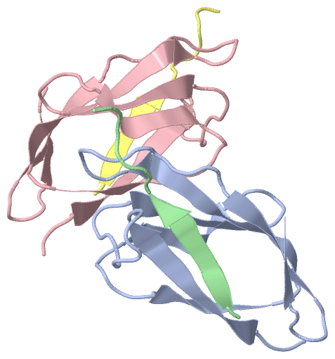
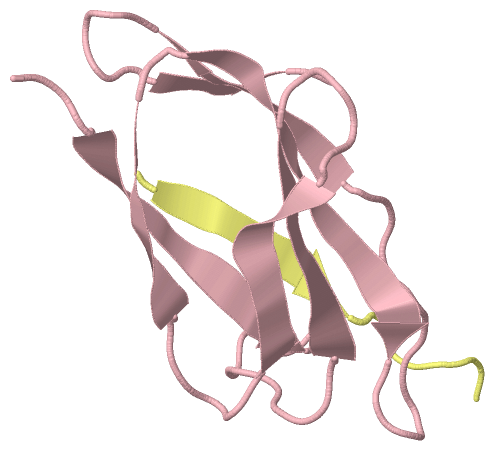
Chain A from PDB  Type:PROTEIN  Length:82
                                                                                                                  
               SCOP domains ---------------------------------------------------------------------------------- SCOP domains
               CATH domains ---------------------------------------------------------------------------------- CATH domains
               Pfam domains ---------------------------------------------------------------------------------- Pfam domains
         Sec.struct. author ...eeeeeeee..........eeeee.....eeeeee.....eeeee..eeeeeeeee..........eeeee.....eee. Sec.struct. author
                 SAPs(SNPs) ---------------------------------------------------------------------------------- SAPs(SNPs)
                    PROSITE ---------------------------------------------------------------------------------- PROSITE
                 Transcript ---------------------------------------------------------------------------------- Transcript
                 4mli A  22 DSATHIKFSKRDEDGKELAGATMELRDSSGKTISTWISDGQVKDFYLYPGKYTFVETAAPDGYEVATAITFTVNEQGQVTVN 103
                                    31        41        51        61        71        81        91       101  

Chain B from PDB  Type:PROTEIN  Length:12
                                            
               SCOP domains ------------ SCOP domains
               CATH domains ------------ CATH domains
               Pfam domains ------------ Pfam domains
         Sec.struct. author .eeeee...... Sec.struct. author
                 SAPs(SNPs) ------------ SAPs(SNPs)
                    PROSITE ------------ PROSITE
                 Transcript ------------ Transcript
                 4mli B 111 AHIVMVDAYKPT 122
                                   120  

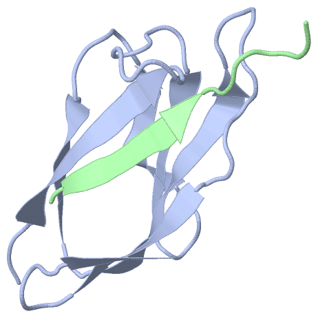
Chain C from PDB  Type:PROTEIN  Length:83
                                                                                                                   
               SCOP domains ----------------------------------------------------------------------------------- SCOP domains
               CATH domains ----------------------------------------------------------------------------------- CATH domains
               Pfam domains ----------------------------------------------------------------------------------- Pfam domains
         Sec.struct. author ...eeeeeeee..........eeeee.....eeeeee.....eeeee..eeeeeeeee..........eeeee.....eee.. Sec.struct. author
                 SAPs(SNPs) ----------------------------------------------------------------------------------- SAPs(SNPs)
                    PROSITE ----------------------------------------------------------------------------------- PROSITE
                 Transcript ----------------------------------------------------------------------------------- Transcript
                 4mli C  22 DSATHIKFSKRDEDGKELAGATMELRDSSGKTISTWISDGQVKDFYLYPGKYTFVETAAPDGYEVATAITFTVNEQGQVTVNG 104
                                    31        41        51        61        71        81        91       101   

Chain D from PDB  Type:PROTEIN  Length:12
                                            
               SCOP domains ------------ SCOP domains
               CATH domains ------------ CATH domains
               Pfam domains ------------ Pfam domains
         Sec.struct. author .eeeee...... Sec.struct. author
                 SAPs(SNPs) ------------ SAPs(SNPs)
                    PROSITE ------------ PROSITE
                 Transcript ------------ Transcript
                 4mli D 111 AHIVMVDAYKPT 122
                                   120  

   Legend:   → Mismatch (orange background)
  - → Gap (green background, '-', border residues have a numbering label)
    → Modified Residue (blue background, lower-case, 'x' indicates undefined single-letter code, labelled with number + name)
  x → Chemical Group (purple background, 'x', labelled with number + name, e.g. ACE or NH2)
  extra numbering lines below/above indicate numbering irregularities and modified residue names etc., number ends below/above '|'

 Classification and Annotation

(-) SCOP Domains  (0, 0)

(no "SCOP Domain" information available for 4MLI)

(-) CATH Domains  (0, 0)

(no "CATH Domain" information available for 4MLI)

(-) Pfam Domains  (0, 0)

(no "Pfam Domain" information available for 4MLI)

(-) Gene Ontology  (0, 0)

Asymmetric Unit(hide GO term definitions)
    (no "Gene Ontology" information available for 4MLI)

 Visualization

(-) Interactive Views

Asymmetric Unit
  Complete Structure
    Jena3D(integrated viewing of ligand, site, SAP, PROSITE, SCOP information)
    WebMol | AstexViewer[tm]@PDBe
(Java Applets, require no local installation except for Java; loading may be slow)
    STRAP
(Java WebStart application, automatic local installation, requires Java; full application with system access!)
    RasMol
(require local installation)
    Molscript (VRML)
(requires installation of a VRML viewer; select preferred view via VRML and generate a mono or stereo PDF format file)
 
  Ligands, Modified Residues, Ions
(no "Ligands, Modified Residues, Ions" information available for 4mli)
 
  Sites
(no "Sites" information available for 4mli)
 
  Cis Peptide Bonds
(no "Cis Peptide Bonds" information available for 4mli)
 
Biological Units
  Complete Structure
    Biological Unit 1  [ Jena3D ]
    Biological Unit 2  [ Jena3D ]

(-) Still Images

Jmol
  protein: cartoon or spacefill or dots and stick; nucleic acid: cartoon and stick; ligands: spacefill; active site: stick
Molscript
  protein, nucleic acid: cartoon; ligands: spacefill; active site: ball and stick

 Databases and Analysis Tools

(-) Databases

Access by PDB/NDB ID
  4mli
    Family and Domain InformationProDom | SYSTERS
    General Structural InformationGlycoscienceDB | MMDB | NDB | OCA | PDB | PDBe | PDBj | PDBsum | PDBWiki | PQS | PROTEOPEDIA
    Orientation in MembranesOPM
    Protein SurfaceSURFACE
    Secondary StructureDSSP (structure derived) | HSSP (homology derived)
    Structural GenomicsGeneCensus
    Structural NeighboursCE | VAST
    Structure ClassificationCATH | Dali | SCOP
    Validation and Original DataBMRB Data View | BMRB Restraints Grid | EDS | PROCHECK | RECOORD | WHAT_CHECK
 
Access by UniProt ID/Accession number
  Q8G9G1_STRPY | Q8G9G1
    Comparative Protein Structure ModelsModBase
    Genomic InformationEnsembl
    Protein-protein InteractionDIP
    Sequence, Family and Domain InformationInterPro | Pfam | SMART | UniProtKB/TrEMBL
 
Access by Enzyme Classificator   (EC Number)
  (no 'Enzyme Classificator' available)
    General Enzyme InformationBRENDA | EC-PDB | Enzyme | IntEnz
    PathwayKEGG | MetaCyc
 
Access by Disease Identifier   (MIM ID)
  (no 'MIM ID' available)
    Disease InformationOMIM
 
Access by GenAge ID
  (no 'GenAge ID' available)
    Age Related InformationGenAge

(-) Analysis Tools

Access by PDB/NDB ID
    Domain InformationXDom
    Interatomic Contacts of Structural UnitsCSU
    Ligand-protein ContactsLPC
    Protein CavitiescastP
    Sequence and Secondary StructurePDBCartoon
    Structure AlignmentSTRAP(Java WebStart application, automatic local installation, requires Java; full application with system access!)
    Structure and Sequence BrowserSTING
 
Access by UniProt ID/Accession number
  Q8G9G1_STRPY | Q8G9G1
    Protein Disorder PredictionDisEMBL | FoldIndex | GLOBPLOT (for more information see DisProt)

 Related Entries

(-) Entries Sharing at Least One Protein Chain (UniProt ID)

UniProtKB/TrEMBL
        Q8G9G1_STRPY | Q8G9G12x5p 4mls

(-) Related Entries Specified in the PDB File

4mls