Show PDB file:   
         Plain Text   HTML   (compressed file size)
QuickSearch:   
by PDB,NDB,UniProt,PROSITE Code or Search Term(s)  
(-)Asymmetric Unit
(-)Asym. Unit - sites
(-)Biological Unit 1
(-)Biol. Unit 1 - sites
(-)Biological Unit 2
collapse expand < >
Image Asymmetric Unit
Asymmetric Unit  (Jmol Viewer)
Image Asym. Unit - sites
Asym. Unit - sites  (Jmol Viewer)
Image Biological Unit 1
Biological Unit 1  (Jmol Viewer)
Image Biol. Unit 1 - sites
Biol. Unit 1 - sites  (Jmol Viewer)
Image Biological Unit 2
Biological Unit 2  (Jmol Viewer)

(-) Description

Title :  CRYSTAL STRUCTURE OF 9,10FN3-ELEGANTIN CHIMERA
 
Authors :  Y. S. Chang, J. H. Shiu, W. J. Chuang
Date :  30 Jul 13  (Deposition) - 06 Aug 14  (Release) - 06 Aug 14  (Revision)
Method :  X-RAY DIFFRACTION
Resolution :  1.42
Chains :  Asym. Unit :  A,B
Biol. Unit 1:  A  (1x)
Biol. Unit 2:  B  (1x)
Keywords :  Fibronectin Type Iii Domain, Cell Binding, Integrin Alpha5Beta1, Cell Adhesion (Keyword Search: [Gene Ontology, PubMed, Web (Google))
 
Reference :  Y. S. Chang, J. H. Shiu, W. J. Chuang
Design, Structure Determination, And Biological Evaluation Of Potent Integrin Alpha5Beta1-Specific Antagonist Using Th Ninth And Tenth Module Of Fibronectin Type Iii Domain
To Be Published
PubMed: search

(-) Compounds

Molecule 1 - FIBRONECTIN
    ChainsA, B
    EngineeredYES
    Expression SystemESCHERICHIA COLI
    Expression System PlasmidPET-21A
    Expression System StrainBL-21
    Expression System Taxid562
    Expression System Vector TypePLASMID
    GeneFN1, FN
    MutationYES
    Organism CommonHUMAN
    Organism ScientificHOMO SAPIENS
    Organism Taxid9606
    SynonymFN, COLD-INSOLUBLE GLOBULIN, CIG, ANASTELLIN, UGL-Y1, UGL- Y2, UGL-Y3

 Structural Features

(-) Chains, Units

  12
Asymmetric Unit AB
Biological Unit 1 (1x)A 
Biological Unit 2 (1x) B

Summary Information (see also Sequences/Alignments below)

(-) Ligands, Modified Residues, Ions  (1, 2)

Asymmetric Unit (1, 2)
No.NameCountTypeFull Name
1CA2Ligand/IonCALCIUM ION
Biological Unit 1 (0, 0)
No.NameCountTypeFull Name
1CA-1Ligand/IonCALCIUM ION
Biological Unit 2 (0, 0)
No.NameCountTypeFull Name
1CA-1Ligand/IonCALCIUM ION

(-) Sites  (2, 2)

Asymmetric Unit (2, 2)
No.NameEvidenceResiduesDescription
1AC1SOFTWAREGLU A:1364 , GLU A:1392 , TRP B:1437 , ASP B:1438 , HOH B:1718 , HOH B:1848BINDING SITE FOR RESIDUE CA A 1601
2AC2SOFTWARETRP A:1437 , ASP A:1438 , HOH A:1705 , HOH A:1708 , GLU B:1364 , GLU B:1392BINDING SITE FOR RESIDUE CA B 1601

(-) SS Bonds  (2, 2)

Asymmetric Unit
No.Residues
1A:1490 -A:1499
2B:1490 -B:1499

(-) Cis Peptide Bonds  (0, 0)

(no "Cis Peptide Bond" information available for 4LXO)

 Sequence-Structure Mapping

(-) SAPs(SNPs)/Variants  (0, 0)

(no "SAP(SNP)/Variant" information available for 4LXO)

(-) PROSITE Motifs  (0, 0)

(no "PROSITE Motif" information available for 4LXO)

(-) Exons   (0, 0)

(no "Exon" information available for 4LXO)

(-) Sequences/Alignments

Asymmetric Unit
   Reformat: Number of residues per line =  ('0' or empty: single-line sequence representation)
  Number of residues per labelling interval =   
  UniProt sequence: complete  aligned part    
   Show mapping: SCOP domains CATH domains Pfam domains Secondary structure (by author)
SAPs(SNPs) PROSITE motifs Exons
(details for a mapped element are shown in a popup box when the mouse pointer rests over it)
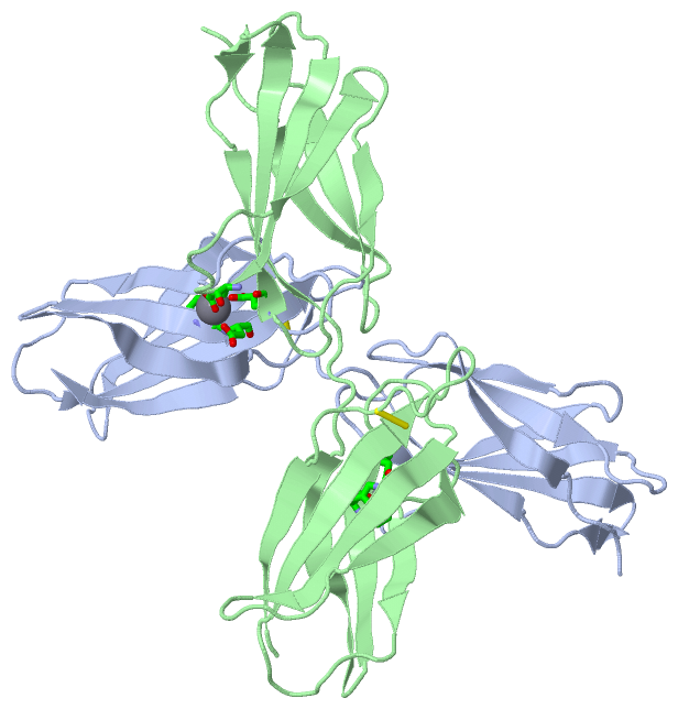
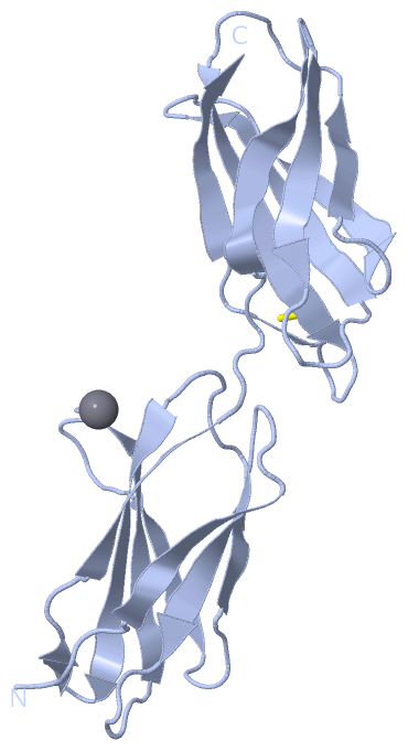
Chain A from PDB  Type:PROTEIN  Length:184
                                                                                                                                                                                                                         
               SCOP domains ---------------------------------------------------------------------------------------------------------------------------------------------------------------------------------------- SCOP domains
               CATH domains ---------------------------------------------------------------------------------------------------------------------------------------------------------------------------------------- CATH domains
               Pfam domains ---------------------------------------------------------------------------------------------------------------------------------------------------------------------------------------- Pfam domains
         Sec.struct. author .....eeeeee......eeeeee.......eeeeeeee.......eeeee.....eeee.......eeeeeeeeee..ee...eeeeee......eeeeeeeee..eeeeeee.......eeeeeeee.......eeeeee....eeee.......eeeeeeeeee......ee...eeeeee. Sec.struct. author
                 SAPs(SNPs) ---------------------------------------------------------------------------------------------------------------------------------------------------------------------------------------- SAPs(SNPs)
                    PROSITE ---------------------------------------------------------------------------------------------------------------------------------------------------------------------------------------- PROSITE
                 Transcript ---------------------------------------------------------------------------------------------------------------------------------------------------------------------------------------- Transcript
                4lxo A 1326 GLDSPTGIDFSDITANSFTVHWIAPRATITGYRIRHHPEHFSGRPREDRVPHSRNSITLTNLTPGTEYVVSIVALNGREESPPLIGQQSTVSDVPRDLEVVAATPTSLLISWDAPAVTVRYYRITYGETGGNSPVQEFTVPGSKSTATISGLKPGVDYTITVYACRARGDNPDCSKPISINYRT 1509
                                  1335      1345      1355      1365      1375      1385      1395      1405      1415      1425      1435      1445      1455      1465      1475      1485      1495      1505    

Chain B from PDB  Type:PROTEIN  Length:184
                                                                                                                                                                                                                         
               SCOP domains ---------------------------------------------------------------------------------------------------------------------------------------------------------------------------------------- SCOP domains
               CATH domains ---------------------------------------------------------------------------------------------------------------------------------------------------------------------------------------- CATH domains
               Pfam domains ---------------------------------------------------------------------------------------------------------------------------------------------------------------------------------------- Pfam domains
         Sec.struct. author .....eeeeee......eeeeee.......eeeeeeee.......eeeeee....eeee.......eeeeeeeeee..ee...eeeeee......eeeeeeeee..eeeeeee.......eeeeeeee.......eeeeee....eeee........eeeeeeee............eeeee.. Sec.struct. author
                 SAPs(SNPs) ---------------------------------------------------------------------------------------------------------------------------------------------------------------------------------------- SAPs(SNPs)
                    PROSITE ---------------------------------------------------------------------------------------------------------------------------------------------------------------------------------------- PROSITE
                 Transcript ---------------------------------------------------------------------------------------------------------------------------------------------------------------------------------------- Transcript
                4lxo B 1326 GLDSPTGIDFSDITANSFTVHWIAPRATITGYRIRHHPEHFSGRPREDRVPHSRNSITLTNLTPGTEYVVSIVALNGREESPPLIGQQSTVSDVPRDLEVVAATPTSLLISWDAPAVTVRYYRITYGETGGNSPVQEFTVPGSKSTATISGLKPGVDYTITVYACRARGDNPDCSKPISINYRT 1509
                                  1335      1345      1355      1365      1375      1385      1395      1405      1415      1425      1435      1445      1455      1465      1475      1485      1495      1505    

   Legend:   → Mismatch (orange background)
  - → Gap (green background, '-', border residues have a numbering label)
    → Modified Residue (blue background, lower-case, 'x' indicates undefined single-letter code, labelled with number + name)
  x → Chemical Group (purple background, 'x', labelled with number + name, e.g. ACE or NH2)
  extra numbering lines below/above indicate numbering irregularities and modified residue names etc., number ends below/above '|'

 Classification and Annotation

(-) SCOP Domains  (0, 0)

(no "SCOP Domain" information available for 4LXO)

(-) CATH Domains  (0, 0)

(no "CATH Domain" information available for 4LXO)

(-) Pfam Domains  (0, 0)

(no "Pfam Domain" information available for 4LXO)

(-) Gene Ontology  (63, 63)

Asymmetric Unit(hide GO term definitions)

 Visualization

(-) Interactive Views

Asymmetric Unit
  Complete Structure
    Jena3D(integrated viewing of ligand, site, SAP, PROSITE, SCOP information)
    WebMol | AstexViewer[tm]@PDBe
(Java Applets, require no local installation except for Java; loading may be slow)
    STRAP
(Java WebStart application, automatic local installation, requires Java; full application with system access!)
    RasMol
(require local installation)
    Molscript (VRML)
(requires installation of a VRML viewer; select preferred view via VRML and generate a mono or stereo PDF format file)
 
  Ligands, Modified Residues, Ions
    CA  [ RasMol | Jena3D ]  +environment [ RasMol | Jena3D ]
 
  Sites
    AC1  [ RasMol ]  +environment [ RasMol ]
    AC2  [ RasMol ]  +environment [ RasMol ]
 
  Cis Peptide Bonds
(no "Cis Peptide Bonds" information available for 4lxo)
 
Biological Units
  Complete Structure
    Biological Unit 1  [ Jena3D ]
    Biological Unit 2  [ Jena3D ]

(-) Still Images

Jmol
  protein: cartoon or spacefill or dots and stick; nucleic acid: cartoon and stick; ligands: spacefill; active site: stick
Molscript
  protein, nucleic acid: cartoon; ligands: spacefill; active site: ball and stick

 Databases and Analysis Tools

(-) Databases

Access by PDB/NDB ID
  4lxo
    Family and Domain InformationProDom | SYSTERS
    General Structural InformationGlycoscienceDB | MMDB | NDB | OCA | PDB | PDBe | PDBj | PDBsum | PDBWiki | PQS | PROTEOPEDIA
    Orientation in MembranesOPM
    Protein SurfaceSURFACE
    Secondary StructureDSSP (structure derived) | HSSP (homology derived)
    Structural GenomicsGeneCensus
    Structural NeighboursCE | VAST
    Structure ClassificationCATH | Dali | SCOP
    Validation and Original DataBMRB Data View | BMRB Restraints Grid | EDS | PROCHECK | RECOORD | WHAT_CHECK
 
Access by UniProt ID/Accession number
  FINC_HUMAN | P02751
    Comparative Protein Structure ModelsModBase
    Genomic InformationEnsembl
    Protein-protein InteractionDIP
    Sequence, Family and Domain InformationInterPro | Pfam | SMART | UniProtKB/SwissProt
 
Access by Enzyme Classificator   (EC Number)
  (no 'Enzyme Classificator' available)
    General Enzyme InformationBRENDA | EC-PDB | Enzyme | IntEnz
    PathwayKEGG | MetaCyc
 
Access by Disease Identifier   (MIM ID)
  (no 'MIM ID' available)
    Disease InformationOMIM
 
Access by GenAge ID
  (no 'GenAge ID' available)
    Age Related InformationGenAge

(-) Analysis Tools

Access by PDB/NDB ID
    Domain InformationXDom
    Interatomic Contacts of Structural UnitsCSU
    Ligand-protein ContactsLPC
    Protein CavitiescastP
    Sequence and Secondary StructurePDBCartoon
    Structure AlignmentSTRAP(Java WebStart application, automatic local installation, requires Java; full application with system access!)
    Structure and Sequence BrowserSTING
 
Access by UniProt ID/Accession number
  FINC_HUMAN | P02751
    Protein Disorder PredictionDisEMBL | FoldIndex | GLOBPLOT (for more information see DisProt)

 Related Entries

(-) Entries Sharing at Least One Protein Chain (UniProt ID)

UniProtKB/Swiss-Prot
        FINC_HUMAN | P027511e88 1e8b 1fbr 1fna 1fnf 1fnh 1j8k 1o9a 1oww 1q38 1qgb 1qo6 1ttf 1ttg 2cg6 2cg7 2ck2 2cku 2ec3 2fn2 2fnb 2gee 2h41 2h45 2ha1 2mnu 2n1k 2ocf 2rky 2rkz 2rl0 3cal 3ejh 3gxe 3m7p 3mql 3r8q 3t1w 3zrz 4gh7 4je4 4jeg 4mmx 4mmy 4mmz 4pz5 5dc0 5dc4 5dc9 5dft 5j6z 5j7c

(-) Related Entries Specified in the PDB File

4lxn