|
|
|
|
 Description
|
|
Compounds
|
||||||||||||||||||||
Chains, Units
Summary Information (see also Sequences/Alignments below) |
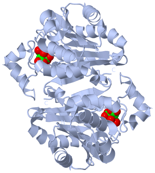
Ligands, Modified Residues, Ions (1, 1)
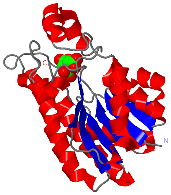
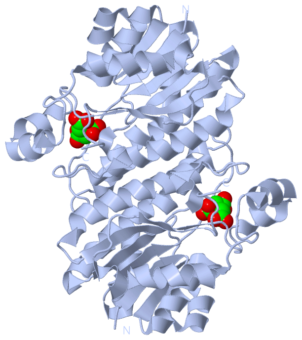
Asymmetric Unit (1, 1)
|
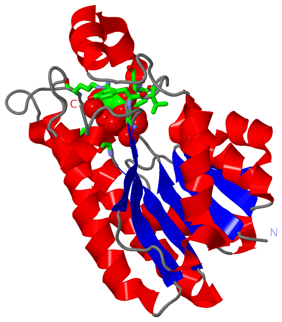
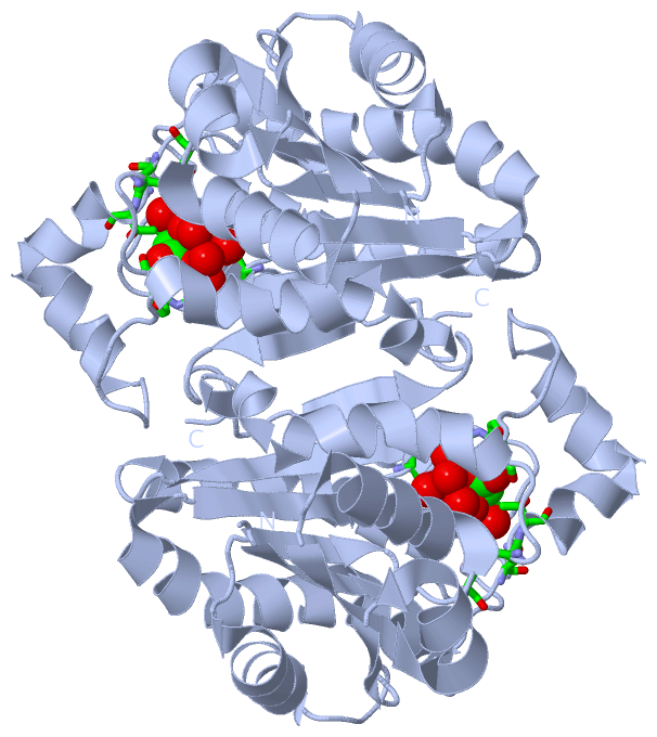
Sites (1, 1)
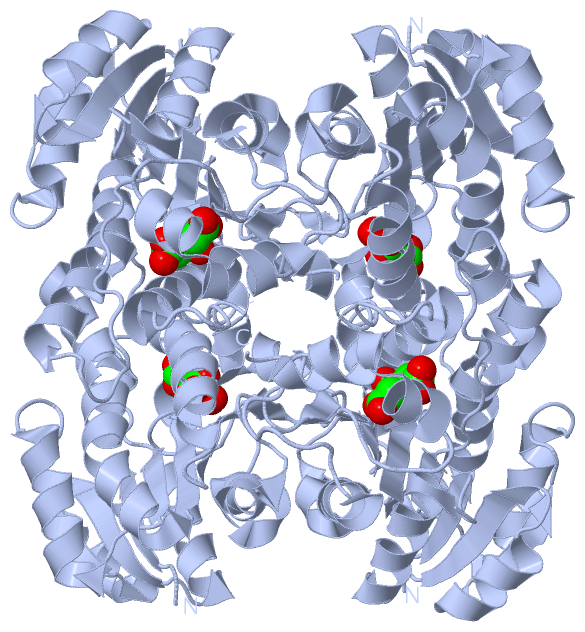
Asymmetric Unit (1, 1)
|
SS Bonds (0, 0)| (no "SS Bond" information available for 4JP3) |
Cis Peptide Bonds (0, 0)| (no "Cis Peptide Bond" information available for 4JP3) |
SAPs(SNPs)/Variants (0, 0)| (no "SAP(SNP)/Variant" information available for 4JP3) |
PROSITE Motifs (0, 0)| (no "PROSITE Motif" information available for 4JP3) |
Exons (0, 0)| (no "Exon" information available for 4JP3) |
Sequences/Alignments
Asymmetric Unit
Chain A from PDB Type:PROTEIN Length:238
SCOP domains d4jp3a_ A: automated matches SCOP domains
CATH domains ---------------------------------------------------------------------------------------------------------------------------------------------------------------------------------------------------------------------------------------------- CATH domains
Pfam domains ---------------------------------------------------------------------------------------------------------------------------------------------------------------------------------------------------------------------------------------------- Pfam domains
SAPs(SNPs) ---------------------------------------------------------------------------------------------------------------------------------------------------------------------------------------------------------------------------------------------- SAPs(SNPs)
PROSITE ---------------------------------------------------------------------------------------------------------------------------------------------------------------------------------------------------------------------------------------------- PROSITE
Transcript ---------------------------------------------------------------------------------------------------------------------------------------------------------------------------------------------------------------------------------------------- Transcript
4jp3 A 2 ERKALVTGGSRGIGRAIAEALVARGYRVAIASRNPEEAAQSLGAVPLPTDLEKDDPKGLVKRALEALGGLHVLVHAAAVNVRKPALELSYEEWRRVLYLHLDVAFLLAQAAAPHMAEAGWGRVLFIGSVTTFTAGGPVPIPAYTTAKTALLGLTRALAKEWARLGIRVNLLCPGYVETEFTLPLRQNPELYEPITARIPMGRWARPEEIARVAAVLCGDEAEYLTGQAVAVDGGFLAY 239
11 21 31 41 51 61 71 81 91 101 111 121 131 141 151 161 171 181 191 201 211 221 231
|
||||||||||||||||||||
SCOP Domains (1, 1)
Asymmetric Unit
 |
CATH Domains (0, 0)| (no "CATH Domain" information available for 4JP3) |
Pfam Domains (0, 0)| (no "Pfam Domain" information available for 4JP3) |
Gene Ontology (1, 1)|
Asymmetric Unit(hide GO term definitions) |
Interactive Views
|
||||||||||||||||||||||||||||||||||||||||||||||||||||||||||||||||||||||||||||||||||||||||||||||||||||||||||||||||||||||||||||||||||||||||||||||||||
Still Images
|
||||||||||||||||
Databases
|
||||||||||||||||||||||||||||||||||||||||||||||||||||||||||||||||||||||||||||||||||||||||||||||||||||||||||||||||||||||||||||||||||||||||||||||||||||||||||||||||
Analysis Tools
|
|||||||||||||||||||||||||||||||||||||||||||||||||||||||||||||
Entries Sharing at Least One Protein Chain (UniProt ID)
Related Entries Specified in the PDB File
|
|