|
|
|
|
 Description
|
|
Compounds
|
||||||||||||||||||||||||||||||||||||||||||||||||||||
Chains, Units
Summary Information (see also Sequences/Alignments below) |
Ligands, Modified Residues, Ions (0, 0)| (no "Ligand,Modified Residues,Ions" information available for 1X1E) |
Sites (0, 0)| (no "Site" information available for 1X1E) |
SS Bonds (0, 0)| (no "SS Bond" information available for 1X1E) |
Cis Peptide Bonds (0, 0)| (no "Cis Peptide Bond" information available for 1X1E) |
SAPs(SNPs)/Variants (0, 0)| (no "SAP(SNP)/Variant" information available for 1X1E) |
PROSITE Motifs (0, 0)| (no "PROSITE Motif" information available for 1X1E) |
Exons (0, 0)| (no "Exon" information available for 1X1E) |
Sequences/Alignments
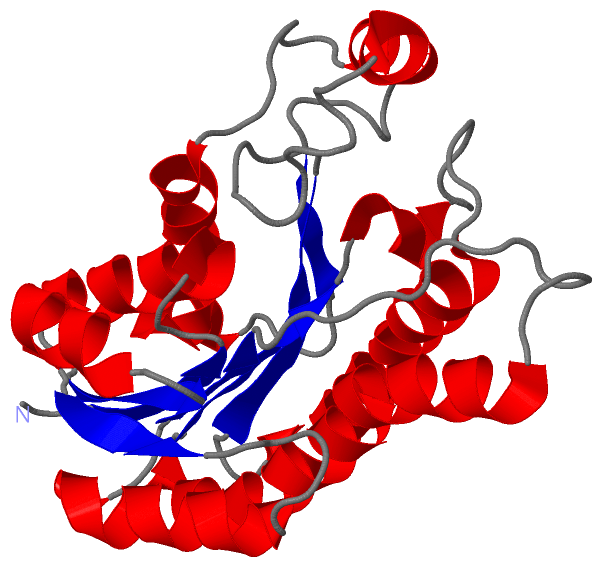
Asymmetric UnitChain A from PDB Type:PROTEIN Length:239 aligned with Q53W82_THET8 | Q53W82 from UniProtKB/TrEMBL Length:239 Alignment length:239 10 20 30 40 50 60 70 80 90 100 110 120 130 140 150 160 170 180 190 200 210 220 230 Q53W82_THET8 1 MERKALVTGGSRGIGRAIAEALVARGYRVAIASRNPEEAAQSLGAVPLPTDLEKDDPKGLVKRALEALGGLHVLVHAAAVNVRKPALELSYEEWRRVLYLHLDVAFLLAQAAAPHMAEAGWGRVLFIGSVTTFTAGGPVPIPAYTTAKTALLGLTRALAKEWARLGIRVNLLCPGYVETEFTLPLRQNPELYEPITARIPMGRWARPEEIARVAAVLCGDEAEYLTGQAVAVDGGFLAY 239 SCOP domains d1x1ea_ A: automated matches SCOP domains CATH domains 1x1eA00 A:1-239 NAD(P)-binding Rossmann-like Domain CATH domains Pfam domains --------adh_short_C2-1x1eA01 A:9-237 -- Pfam domains SAPs(SNPs) ----------------------------------------------------------------------------------------------------------------------------------------------------------------------------------------------------------------------------------------------- SAPs(SNPs) PROSITE ----------------------------------------------------------------------------------------------------------------------------------------------------------------------------------------------------------------------------------------------- PROSITE Transcript ----------------------------------------------------------------------------------------------------------------------------------------------------------------------------------------------------------------------------------------------- Transcript 1x1e A 1 MERKALVTGGSRGIGRAIAEALVARGYRVAIASRNPEEAAQSLGAVPLPTDLEKDDPKGLVKRALEALGGLHVLVHAAAVNVRKPALELSYEEWRRVLYLHLDVAFLLAQAAAPHMAEAGWGRVLFIGSVTTFTAGGPVPIPAYTTAKTALLGLTRALAKEWARLGIRVNLLCPGYVETEFTLPLRQNPELYEPITARIPMGRWARPEEIARVAAVLCGDEAEYLTGQAVAVDGGFLAY 239 10 20 30 40 50 60 70 80 90 100 110 120 130 140 150 160 170 180 190 200 210 220 230
|
||||||||||||||||||||
SCOP Domains (1, 1)
Asymmetric Unit
 |
CATH Domains (1, 1)
Asymmetric Unit
|
Pfam Domains (1, 1)
Asymmetric Unit
|
Gene Ontology (1, 1)|
Asymmetric Unit(hide GO term definitions) Chain A (Q53W82_THET8 | Q53W82)
|
||||||||||||
Interactive Views
|
||||||||||||||||||||||||||||||||||||||||||||||||||||||||||||||||||||||||||||||||||||||||||||||||||||||||||||||||||||||||||||||||||||||
Still Images
|
||||||||||||||||
Databases
|
||||||||||||||||||||||||||||||||||||||||||||||||||||||||||||||||||||||||||||||||||||||||||||||||||||||||||||||||||||||||||||||||||||||||||||||||||||||||||||||||
Analysis Tools
|
|||||||||||||||||||||||||||||||||||||||||||||||||||||||||||||
Entries Sharing at Least One Protein Chain (UniProt ID)
Related Entries Specified in the PDB File
|
|