|
|
|
|
 Description
|
|
Compounds
|
||||||||||||||||||||||||||||||||||||||||||||||||||||||||
Chains, Units
Summary Information (see also Sequences/Alignments below) |
Ligands, Modified Residues, Ions (0, 0)| (no "Ligand,Modified Residues,Ions" information available for 4J5O) |
Sites (0, 0)| (no "Site" information available for 4J5O) |
SS Bonds (0, 0)| (no "SS Bond" information available for 4J5O) |
Cis Peptide Bonds (0, 0)| (no "Cis Peptide Bond" information available for 4J5O) |
SAPs(SNPs)/Variants (0, 0)| (no "SAP(SNP)/Variant" information available for 4J5O) |
PROSITE Motifs (0, 0)| (no "PROSITE Motif" information available for 4J5O) |
Exons (0, 0)| (no "Exon" information available for 4J5O) |
Sequences/Alignments
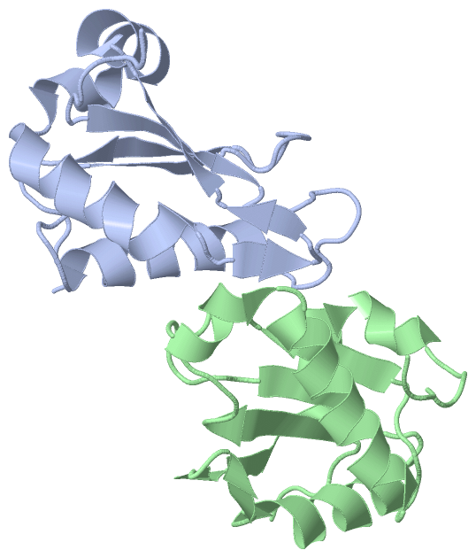
Asymmetric Unit
Chain A from PDB Type:PROTEIN Length:111
SCOP domains --------------------------------------------------------------------------------------------------------------- SCOP domains
CATH domains --------------------------------------------------------------------------------------------------------------- CATH domains
Pfam domains --------------------------------------------------------------------------------------------------------------- Pfam domains
SAPs(SNPs) --------------------------------------------------------------------------------------------------------------- SAPs(SNPs)
PROSITE --------------------------------------------------------------------------------------------------------------- PROSITE
Transcript --------------------------------------------------------------------------------------------------------------- Transcript
4j5o A 418 VQPLATQCFQLSNMFNPQTEEEVGWDTEIKDDVIEECNKHGGVIHIYVDKNSAQGNVYVKCPSIAAAIAAVNALHGRWFAGKMITAAYVPLPTYHNLFPDSMTATQLLVPS 528
427 437 447 457 467 477 487 497 507 517 527
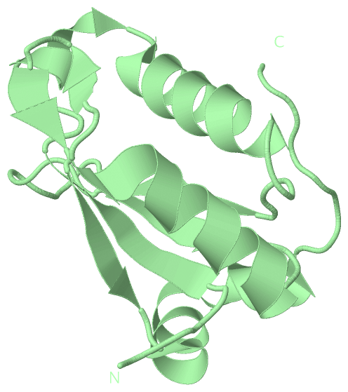
Chain B from PDB Type:PROTEIN Length:109
SCOP domains ------------------------------------------------------------------------------------------------------------- SCOP domains
CATH domains ------------------------------------------------------------------------------------------------------------- CATH domains
Pfam domains ------------------------------------------------------------------------------------------------------------- Pfam domains
SAPs(SNPs) ------------------------------------------------------------------------------------------------------------- SAPs(SNPs)
PROSITE ------------------------------------------------------------------------------------------------------------- PROSITE
Transcript ------------------------------------------------------------------------------------------------------------- Transcript
4j5o B 420 PLATQCFQLSNMFNPQTEEEVGWDTEIKDDVIEECNKHGGVIHIYVDKNSAQGNVYVKCPSIAAAIAAVNALHGRWFAGKMITAAYVPLPTYHNLFPDSMTATQLLVPS 528
429 439 449 459 469 479 489 499 509 519
|
||||||||||||||||||||
SCOP Domains (0, 0)| (no "SCOP Domain" information available for 4J5O) |
CATH Domains (0, 0)| (no "CATH Domain" information available for 4J5O) |
Pfam Domains (0, 0)| (no "Pfam Domain" information available for 4J5O) |
Gene Ontology (13, 13)|
Asymmetric Unit(hide GO term definitions) |
Interactive Views
|
|||||||||||||||||||||||||||||||||||||||||||||||||||||||||||||||||||||||||||||||||||||||||||||||||||||||||||||||||||||||||||||||||||||||||||
Still Images
|
||||||||||||||||
Databases
|
||||||||||||||||||||||||||||||||||||||||||||||||||||||||||||||||||||||||||||||||||||||||||||||||||||||||||||||||||||||||||||||||||||||||||||||||||||||||||||||||
Analysis Tools
|
|||||||||||||||||||||||||||||||||||||||||||||||||||||||||||||
Entries Sharing at Least One Protein Chain (UniProt ID)
Related Entries Specified in the PDB File
|
|