Show PDB file:   
         Plain Text   HTML   (compressed file size)
QuickSearch:   
by PDB,NDB,UniProt,PROSITE Code or Search Term(s)  
(-)Asymmetric Unit
(-)Asym. Unit - sites
(-)Biological Unit 1
(-)Biol. Unit 1 - sites
(-)Biological Unit 2
collapse expand < >
Image Asymmetric Unit
Asymmetric Unit  (Jmol Viewer)
Image Asym. Unit - sites
Asym. Unit - sites  (Jmol Viewer)
Image Biological Unit 1
Biological Unit 1  (Jmol Viewer)
Image Biol. Unit 1 - sites
Biol. Unit 1 - sites  (Jmol Viewer)
Image Biological Unit 2
Biological Unit 2  (Jmol Viewer)

(-) Description

Title :  CRYSTAL STRUCTURE OF YELLOWTAIL ASCITES VIRUS VP4 PROTEASE ACTIVE SITE MUTANT (K674A) REVEALS BOTH AN ACYL-ENZYME COMPLEX AND AN EMPTY ACTIVE SITE
 
Authors :  M. Paetzel, I. Y. W. Chung
Date :  30 Jan 13  (Deposition) - 27 Feb 13  (Release) - 22 May 13  (Revision)
Method :  X-RAY DIFFRACTION
Resolution :  2.30
Chains :  Asym. Unit :  A,B
Biol. Unit 1:  A  (1x)
Biol. Unit 2:  B  (1x)
Keywords :  Viral Protease, Birnavirus, Serine-Lysine Dyad Mechanism, Alpha-Beta Protein Fold, Lysine General Base, Acyl-Enzyme, Product Complex, Hydrolase (Keyword Search: [Gene Ontology, PubMed, Web (Google))
 
Reference :  I. Y. Chung, M. Paetzel
Crystal Structures Of Yellowtail Ascites Virus Vp4 Protease Trapping An Internal Cleavage Site Trans Acyl-Enzyme Comple In A Native Ser/Lys Dyad Active Site.
J. Biol. Chem. V. 288 13068 2013
PubMed-ID: 23511637  |  Reference-DOI: 10.1074/JBC.M112.386953

(-) Compounds

Molecule 1 - YELLOWTAIL ASCITES VIRUS (YAV) VP4 PROTEASE
    ChainsA
    EC Number3.4.21.115
    EngineeredYES
    Expression SystemESCHERICHIA COLI
    Expression System PlasmidPET28B+
    Expression System StrainTUNER (DE3)
    Expression System Taxid469008
    Expression System Vector TypePLASMID
    GeneVIRAL PROTEIN 4 (VP4)
    MutationYES
    Organism ScientificYELLOWTAIL ASCITES VIRUS
    Organism Taxid360000
    StrainY-6
 
Molecule 2 - YELLOWTAIL ASCITES VIRUS (YAV) VP4 PROTEASE
    ChainsB
    EC Number3.4.21.115
    EngineeredYES
    Expression SystemESCHERICHIA COLI
    Expression System PlasmidPET28B+
    Expression System StrainTUNER (DE3)
    Expression System Taxid469008
    Expression System Vector TypePLASMID
    GeneVIRAL PROTEIN 4 (VP4)
    MutationYES
    Organism ScientificYELLOWTAIL ASCITES VIRUS
    Organism Taxid360000
    StrainY-6

 Structural Features

(-) Chains, Units

  12
Asymmetric Unit AB
Biological Unit 1 (1x)A 
Biological Unit 2 (1x) B

Summary Information (see also Sequences/Alignments below)

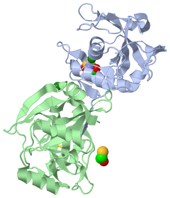
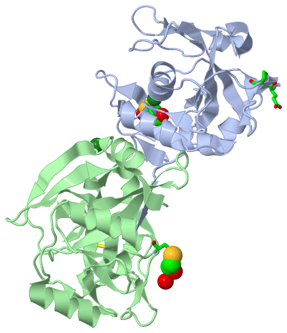
(-) Ligands, Modified Residues, Ions  (3, 4)

Asymmetric Unit (3, 4)
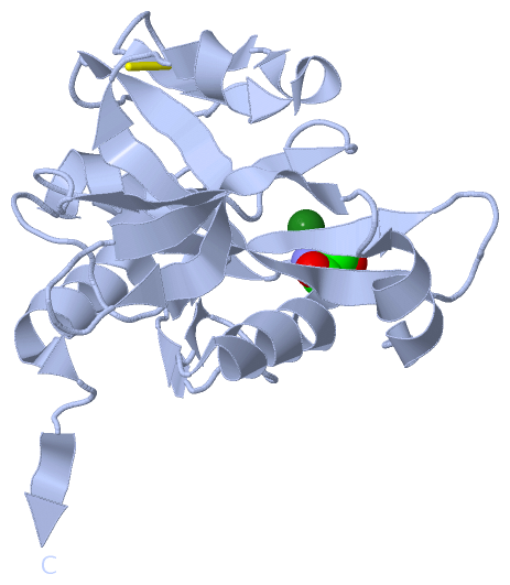
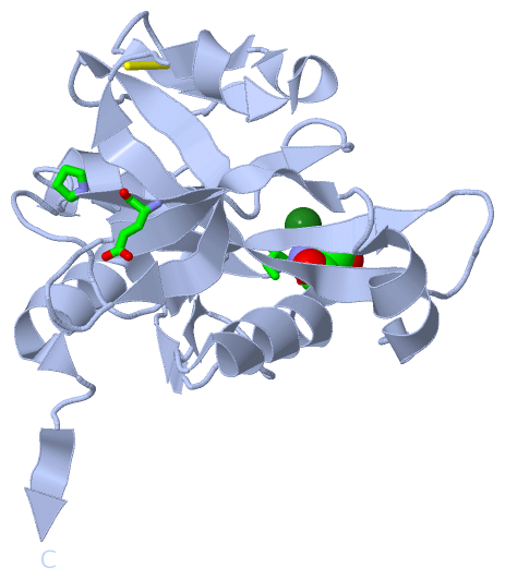
No.NameCountTypeFull Name
1BME1Ligand/IonBETA-MERCAPTOETHANOL
2CSO1Mod. Amino AcidS-HYDROXYCYSTEINE
3MG2Ligand/IonMAGNESIUM ION
Biological Unit 1 (1, 1)
No.NameCountTypeFull Name
1BME-1Ligand/IonBETA-MERCAPTOETHANOL
2CSO1Mod. Amino AcidS-HYDROXYCYSTEINE
3MG-1Ligand/IonMAGNESIUM ION
Biological Unit 2 (1, 1)
No.NameCountTypeFull Name
1BME1Ligand/IonBETA-MERCAPTOETHANOL
2CSO-1Mod. Amino AcidS-HYDROXYCYSTEINE
3MG-1Ligand/IonMAGNESIUM ION

(-) Sites  (3, 3)

Asymmetric Unit (3, 3)
No.NameEvidenceResiduesDescription
1AC1SOFTWAREILE A:668 , CSO A:669BINDING SITE FOR RESIDUE MG A 801
2AC2SOFTWAREGLU A:533 , PRO A:535 , CYS B:669 , HOH B:1069BINDING SITE FOR RESIDUE BME B 801
3AC3SOFTWAREHOH B:1037 , HOH B:1038 , HOH B:1039 , HOH B:1040 , HOH B:1073 , HOH B:1074BINDING SITE FOR RESIDUE MG B 802

(-) SS Bonds  (2, 2)

Asymmetric Unit
No.Residues
1A:588 -A:604
2B:588 -B:604

(-) Cis Peptide Bonds  (0, 0)

(no "Cis Peptide Bond" information available for 4IZK)

 Sequence-Structure Mapping

(-) SAPs(SNPs)/Variants  (0, 0)

(no "SAP(SNP)/Variant" information available for 4IZK)

(-) PROSITE Motifs  (0, 0)

(no "PROSITE Motif" information available for 4IZK)

(-) Exons   (0, 0)

(no "Exon" information available for 4IZK)

(-) Sequences/Alignments

Asymmetric Unit
   Reformat: Number of residues per line =  ('0' or empty: single-line sequence representation)
  Number of residues per labelling interval =   
  UniProt sequence: complete  aligned part    
   Show mapping: SCOP domains CATH domains Pfam domains Secondary structure (by author)
SAPs(SNPs) PROSITE motifs Exons
(details for a mapped element are shown in a popup box when the mouse pointer rests over it)
Chain A from PDB  Type:PROTEIN  Length:202
                                                                                                                                                                                                                                          
               SCOP domains ---------------------------------------------------------------------------------------------------------------------------------------------------------------------------------------------------------- SCOP domains
               CATH domains ---------------------------------------------------------------------------------------------------------------------------------------------------------------------------------------------------------- CATH domains
               Pfam domains ---------------------------------------------------------------------------------------------------------------------------------------------------------------------------------------------------------- Pfam domains
         Sec.struct. author ..hhhhhhhh...eeeeeee......eeeeeeee.......eeeeeeeeee..hhhhhhhhhh.eee.....eeee............eeeeeeeeeee.....eeeeee.........hhhhhhhhhhh........ee..ee......ee...hhhhhhhhhhhh...eee......ee..hhhhhhhhhhhh...eee. Sec.struct. author
                 SAPs(SNPs) ---------------------------------------------------------------------------------------------------------------------------------------------------------------------------------------------------------- SAPs(SNPs)
                    PROSITE ---------------------------------------------------------------------------------------------------------------------------------------------------------------------------------------------------------- PROSITE
                 Transcript ---------------------------------------------------------------------------------------------------------------------------------------------------------------------------------------------------------- Transcript
                 4izk A 515 RFSRNLKDRLESNNYEEMELPPPTKGVIIPVVHTVESAPGEAFGSLLVIIPGAYPELLDPNQQVLSHFKNDTGCVWGIGEDIPFEGDDICYTALPLKEIKKNGNIVVEKVFAGPAMGPSCQLGLSLLVNDIDKGVPRMVFTGEIANDEETIVPIcGVDIAAIAAHEHGLPLVGCQPGVDEVVANTSLASHLIQSGALPVQKA 716
                                   524       534       544       554       564       574       584       594       604       614       624       634       644       654       664    |  674       684       694       704       714  
                                                                                                                                                                                    669-CSO                                           

Chain B from PDB  Type:PROTEIN  Length:203
                                                                                                                                                                                                                                           
               SCOP domains ----------------------------------------------------------------------------------------------------------------------------------------------------------------------------------------------------------- SCOP domains
               CATH domains ----------------------------------------------------------------------------------------------------------------------------------------------------------------------------------------------------------- CATH domains
               Pfam domains ----------------------------------------------------------------------------------------------------------------------------------------------------------------------------------------------------------- Pfam domains
         Sec.struct. author .eehhhhhhhhhh..eeeeee......eeeeeeee......eeeeeeeeeee..hhhhhhhhhh.eee.....eeee............eeeeeeeeeee.....eeeee.....ee...hhhhhhhhhhh........ee..ee......ee...hhhhhhhhhhhh...eee......ee..hhhhhhhhhhhh....... Sec.struct. author
                 SAPs(SNPs) ----------------------------------------------------------------------------------------------------------------------------------------------------------------------------------------------------------- SAPs(SNPs)
                    PROSITE ----------------------------------------------------------------------------------------------------------------------------------------------------------------------------------------------------------- PROSITE
                 Transcript ----------------------------------------------------------------------------------------------------------------------------------------------------------------------------------------------------------- Transcript
                 4izk B 514 GRFSRNLKDRLESNNYEEMELPPPTKGVIIPVVHTVESAPGEAFGSLLVIIPGAYPELLDPNQQVLSHFKNDTGCVWGIGEDIPFEGDDICYTALPLKEIKKNGNIVVEKVFAGPAMGPSCQLGLSLLVNDIDKGVPRMVFTGEIANDEETIVPICGVDIAAIAAHEHGLPLVGCQPGVDEVVANTSLASHLIQSGALPVQKA 716
                                   523       533       543       553       563       573       583       593       603       613       623       633       643       653       663       673       683       693       703       713   

   Legend:   → Mismatch (orange background)
  - → Gap (green background, '-', border residues have a numbering label)
    → Modified Residue (blue background, lower-case, 'x' indicates undefined single-letter code, labelled with number + name)
  x → Chemical Group (purple background, 'x', labelled with number + name, e.g. ACE or NH2)
  extra numbering lines below/above indicate numbering irregularities and modified residue names etc., number ends below/above '|'

 Classification and Annotation

(-) SCOP Domains  (0, 0)

(no "SCOP Domain" information available for 4IZK)

(-) CATH Domains  (0, 0)

(no "CATH Domain" information available for 4IZK)

(-) Pfam Domains  (0, 0)

(no "Pfam Domain" information available for 4IZK)

(-) Gene Ontology  (9, 9)

Asymmetric Unit(hide GO term definitions)

 Visualization

(-) Interactive Views

Asymmetric Unit
  Complete Structure
    Jena3D(integrated viewing of ligand, site, SAP, PROSITE, SCOP information)
    WebMol | AstexViewer[tm]@PDBe
(Java Applets, require no local installation except for Java; loading may be slow)
    STRAP
(Java WebStart application, automatic local installation, requires Java; full application with system access!)
    RasMol
(require local installation)
    Molscript (VRML)
(requires installation of a VRML viewer; select preferred view via VRML and generate a mono or stereo PDF format file)
 
  Ligands, Modified Residues, Ions
    BME  [ RasMol | Jena3D ]  +environment [ RasMol | Jena3D ]
    CSO  [ RasMol | Jena3D ]  +environment [ RasMol | Jena3D ]
    MG  [ RasMol | Jena3D ]  +environment [ RasMol | Jena3D ]
 
  Sites
    AC1  [ RasMol ]  +environment [ RasMol ]
    AC2  [ RasMol ]  +environment [ RasMol ]
    AC3  [ RasMol ]  +environment [ RasMol ]
 
  Cis Peptide Bonds
(no "Cis Peptide Bonds" information available for 4izk)
 
Biological Units
  Complete Structure
    Biological Unit 1  [ Jena3D ]
    Biological Unit 2  [ Jena3D ]

(-) Still Images

Jmol
  protein: cartoon or spacefill or dots and stick; nucleic acid: cartoon and stick; ligands: spacefill; active site: stick
Molscript
  protein, nucleic acid: cartoon; ligands: spacefill; active site: ball and stick

 Databases and Analysis Tools

(-) Databases

Access by PDB/NDB ID
  4izk
    Family and Domain InformationProDom | SYSTERS
    General Structural InformationGlycoscienceDB | MMDB | NDB | OCA | PDB | PDBe | PDBj | PDBsum | PDBWiki | PQS | PROTEOPEDIA
    Orientation in MembranesOPM
    Protein SurfaceSURFACE
    Secondary StructureDSSP (structure derived) | HSSP (homology derived)
    Structural GenomicsGeneCensus
    Structural NeighboursCE | VAST
    Structure ClassificationCATH | Dali | SCOP
    Validation and Original DataBMRB Data View | BMRB Restraints Grid | EDS | PROCHECK | RECOORD | WHAT_CHECK
 
Access by UniProt ID/Accession number
  P89521_9VIRU | P89521
    Comparative Protein Structure ModelsModBase
    Genomic InformationEnsembl
    Protein-protein InteractionDIP
    Sequence, Family and Domain InformationInterPro | Pfam | SMART | UniProtKB/TrEMBL
 
Access by Enzyme Classificator   (EC Number)
  3.4.21.115
    General Enzyme InformationBRENDA | EC-PDB | Enzyme | IntEnz
    PathwayKEGG | MetaCyc
 
Access by Disease Identifier   (MIM ID)
  (no 'MIM ID' available)
    Disease InformationOMIM
 
Access by GenAge ID
  (no 'GenAge ID' available)
    Age Related InformationGenAge

(-) Analysis Tools

Access by PDB/NDB ID
    Domain InformationXDom
    Interatomic Contacts of Structural UnitsCSU
    Ligand-protein ContactsLPC
    Protein CavitiescastP
    Sequence and Secondary StructurePDBCartoon
    Structure AlignmentSTRAP(Java WebStart application, automatic local installation, requires Java; full application with system access!)
    Structure and Sequence BrowserSTING
 
Access by UniProt ID/Accession number
  P89521_9VIRU | P89521
    Protein Disorder PredictionDisEMBL | FoldIndex | GLOBPLOT (for more information see DisProt)

 Related Entries

(-) Entries Sharing at Least One Protein Chain (UniProt ID)

UniProtKB/TrEMBL
        P89521_9VIRU | P895214izj

(-) Related Entries Specified in the PDB File

2gef CRYSTAL STRUCTURE OF VP4 PROTEASE FROM BLOTCHED SNAKEHEAD VIRUS (BSNV)
2pnl CRYSTAL STRUCTURE OF VP4 PROTEASE FROM INFECTIOUS PANCREATIC NECROSIS VIRUS (IPNV) IN SPACE GROUP P1
2pnm CRYSTAL STRUCTURE OF VP4 PROTEASE FROM INFECTIOUS PANCREATIC NECROSIS VIRUS (IPNV) IN SPACE GROUP P6122
3p06 CRYSTAL STRUCTURE OF VP4 PROTEASE FROM TELLINA VIRUS 1 (TV- 1)
4izj