|
|
|
|
 Description
|
|
Compounds
|
||||||||||||||||||||||||||||||||||||||||||||||||||||
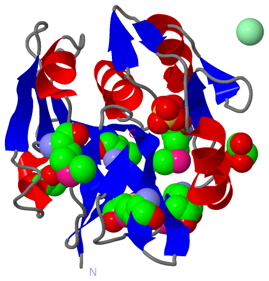
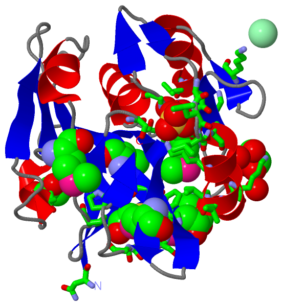
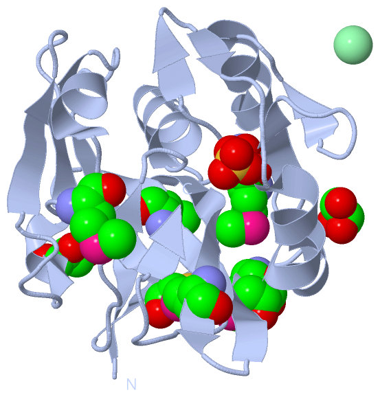
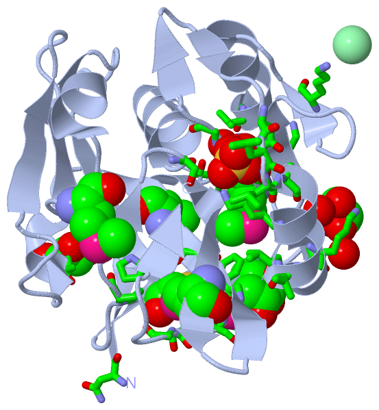
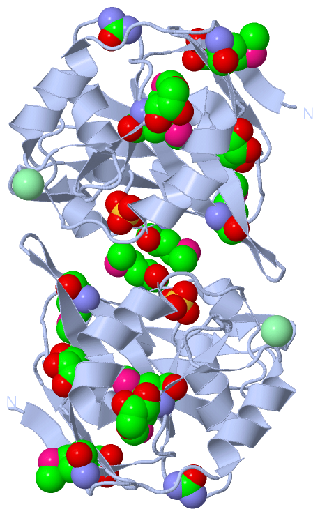
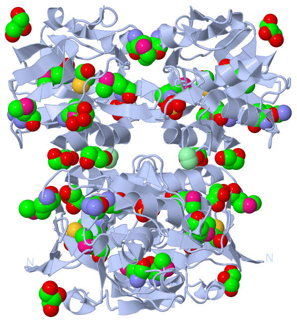
Chains, Units
Summary Information (see also Sequences/Alignments below) |
Ligands, Modified Residues, Ions (6, 12)
Asymmetric Unit (6, 12)
|
Sites (7, 7)
Asymmetric Unit (7, 7)
|
SS Bonds (0, 0)| (no "SS Bond" information available for 3P06) |
Cis Peptide Bonds (0, 0)| (no "Cis Peptide Bond" information available for 3P06) |
SAPs(SNPs)/Variants (0, 0)| (no "SAP(SNP)/Variant" information available for 3P06) |
PROSITE Motifs (0, 0)| (no "PROSITE Motif" information available for 3P06) |
Exons (0, 0)| (no "Exon" information available for 3P06) |
Sequences/Alignments
Asymmetric UnitChain A from PDB Type:PROTEIN Length:194 aligned with Q2PBR5_9VIRU | Q2PBR5 from UniProtKB/TrEMBL Length:1114 Alignment length:194 646 656 666 676 686 696 706 716 726 736 746 756 766 776 786 796 806 816 826 Q2PBR5_9VIRU 637 NGVELSAVGVLLPVLMDSGRRISGGAFMAVKGDLSEHIKNPKNTRIAQTVAGGTIYGLSEMVNIDEAEKLPIKGAITVLPVVQATATSILVPDNQPQLAFNSWEAAACAADTLESQQTPFLMVTGAVESGNLSPNLLAVQKQLLVAKPAGIGLAANSDRALKVVTLEQLRQVVGDKPWRKPMVTFSSGKNVAQA 830 SCOP domains -------------------------------------------------------------------------------------------------------------------------------------------------------------------------------------------------- SCOP domains CATH domains -------------------------------------------------------------------------------------------------------------------------------------------------------------------------------------------------- CATH domains Pfam domains -------------------------------------------------------------------------------------------------------------------------------------------------------------------------------------------------- Pfam domains SAPs(SNPs) -------------------------------------------------------------------------------------------------------------------------------------------------------------------------------------------------- SAPs(SNPs) PROSITE -------------------------------------------------------------------------------------------------------------------------------------------------------------------------------------------------- PROSITE Transcript -------------------------------------------------------------------------------------------------------------------------------------------------------------------------------------------------- Transcript 3p06 A 637 NGVELSAVGVLLPVLmDSGRRISGGAFmAVKGDLSEHIKNPKNTRIAQTVAGGTIYGLSEmVNIDEAEKLPIKGAITVLPVVQATATSILVPDNQPQLAFNSWEAAACAADTLESQQTPFLmVTGAVESGNLSPNLLAVQKQLLVAKPAGIGLAANSDRALKVVTLEQLRQVVGDKPWRKPmVTFSSGKNVAQA 830 646 | 656 666 676 686 696| 706 716 726 736 746 756 | 766 776 786 796 806 816 | 826 652-MSE 664-MSE 697-MSE 758-MSE 818-MSE
|
||||||||||||||||||||
SCOP Domains (0, 0)| (no "SCOP Domain" information available for 3P06) |
CATH Domains (0, 0)| (no "CATH Domain" information available for 3P06) |
Pfam Domains (0, 0)| (no "Pfam Domain" information available for 3P06) |
Gene Ontology (1, 1)|
Asymmetric Unit(hide GO term definitions) Chain A (Q2PBR5_9VIRU | Q2PBR5)
|
||||||||||||
Interactive Views
|
|||||||||||||||||||||||||||||||||||||||||||||||||||||||||||||||||||||||||||||||||||||||||||||||||||||||||||||||||||||||||||||||||||||||||||||||||||||||||||||||||||||||||||||||||||||||||||||||||||||||||||||||||||||||||||||||
Still Images
|
||||||||||||||||
Databases
|
||||||||||||||||||||||||||||||||||||||||||||||||||||||||||||||||||||||||||||||||||||||||||||||||||||||||||||||||||||||||||||||||||||||||||||||||||||||||||||||||
Analysis Tools
|
|||||||||||||||||||||||||||||||||||||||||||||||||||||||||||||
Entries Sharing at Least One Protein Chain (UniProt ID)
Related Entries Specified in the PDB File
|
|