|
|
|
|
 Description
|
|
Compounds
|
||||||||||||||||||||||||||||||||||||||||||||
Chains, Units
Summary Information (see also Sequences/Alignments below) |
Ligands, Modified Residues, Ions (3, 23)| Asymmetric Unit (3, 23) Biological Unit 1 (3, 46) |
Sites (12, 12)
Asymmetric Unit (12, 12)
|
SS Bonds (0, 0)| (no "SS Bond" information available for 4I19) |
Cis Peptide Bonds (1, 1)
Asymmetric Unit
|
||||||||
SAPs(SNPs)/Variants (0, 0)| (no "SAP(SNP)/Variant" information available for 4I19) |
PROSITE Motifs (0, 0)| (no "PROSITE Motif" information available for 4I19) |
Exons (0, 0)| (no "Exon" information available for 4I19) |
Sequences/Alignments
Asymmetric Unit
Chain A from PDB Type:PROTEIN Length:386
SCOP domains -------------------------------------------------------------------------------------------------------------------------------------------------------------------------------------------------------------------------------------------------------------------------------------------------------------------------------------------------------------------------------------------------- SCOP domains
CATH domains -------------------------------------------------------------------------------------------------------------------------------------------------------------------------------------------------------------------------------------------------------------------------------------------------------------------------------------------------------------------------------------------------- CATH domains
Pfam domains -------------------------------------------------------------------------------------------------------------------------------------------------------------------------------------------------------------------------------------------------------------------------------------------------------------------------------------------------------------------------------------------------- Pfam domains
SAPs(SNPs) -------------------------------------------------------------------------------------------------------------------------------------------------------------------------------------------------------------------------------------------------------------------------------------------------------------------------------------------------------------------------------------------------- SAPs(SNPs)
PROSITE -------------------------------------------------------------------------------------------------------------------------------------------------------------------------------------------------------------------------------------------------------------------------------------------------------------------------------------------------------------------------------------------------- PROSITE
Transcript -------------------------------------------------------------------------------------------------------------------------------------------------------------------------------------------------------------------------------------------------------------------------------------------------------------------------------------------------------------------------------------------------- Transcript
4i19 A 0 AmRPFQVQIPQADIDDLKRRLSETRWPELVDVGWSRGAPLSYIKELAEYWRDGFDWRAAERRINQYPQFTTEIDGATIHFLHVRSPEPDATPmVITHGWPGTPVEFLDIIGPLTDPRAHGGDPADAFHLVIPSLPGFGLSGPLKSAGWELGRIAmAWSKLmASLGYERYIAQGGDIGAFTSLLLGAIDPSHLAGIHVNLLQTNLSGEPGELETLSDADKARLAVSERFLDDLSGPmKmQSTRPHTIGYmLNDSPVAQLAYLLEmFKHWAQTENVPEDAVDRDLmLTHISLFWFTATGGSAAQAHYELKPFLPITSLIGRSPTLDVPmGVAVYPGALFQPVRSLAERDFKQIVHWAELDRGGHFSAmEEPDLFVDDLRTFNRTLKKL 385
| 9 19 29 39 49 59 69 79 89 | 99 109 119 129 139 149 | 159| 169 179 189 199 209 219 229 | 239 249 259 | 269 279 | 289 299 309 319 |329 339 349 359 | 369 379
| 92-MSE 154-MSE | 235-MSE 248-MSE 263-MSE 283-MSE 326-MSE 365-MSE
1-MSE 160-MSE 237-MSE
|
||||||||||||||||||||
SCOP Domains (0, 0)| (no "SCOP Domain" information available for 4I19) |
CATH Domains (0, 0)| (no "CATH Domain" information available for 4I19) |
Pfam Domains (0, 0)| (no "Pfam Domain" information available for 4I19) |
Gene Ontology (3, 3)|
Asymmetric Unit(hide GO term definitions) |
Interactive Views
|
||||||||||||||||||||||||||||||||||||||||||||||||||||||||||||||||||||||||||||||||||||||||||||||||||||||||||||||||||||||||||||||||||||||||||||||||||||||||||||||||||||||||||||||||||||||||||||||||||||||||||||||||||||||||||||||||||||
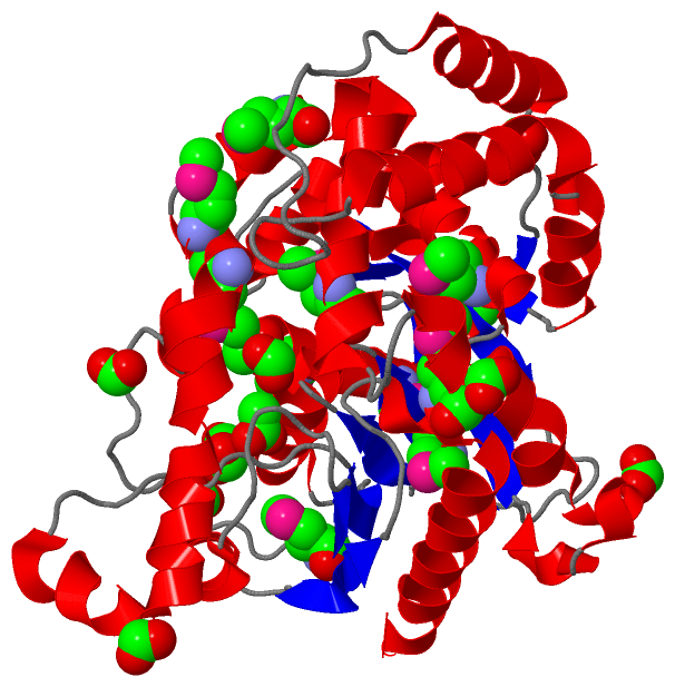
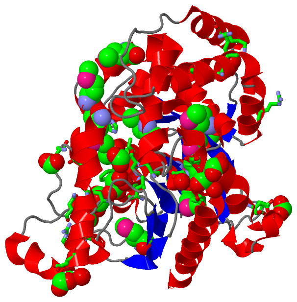
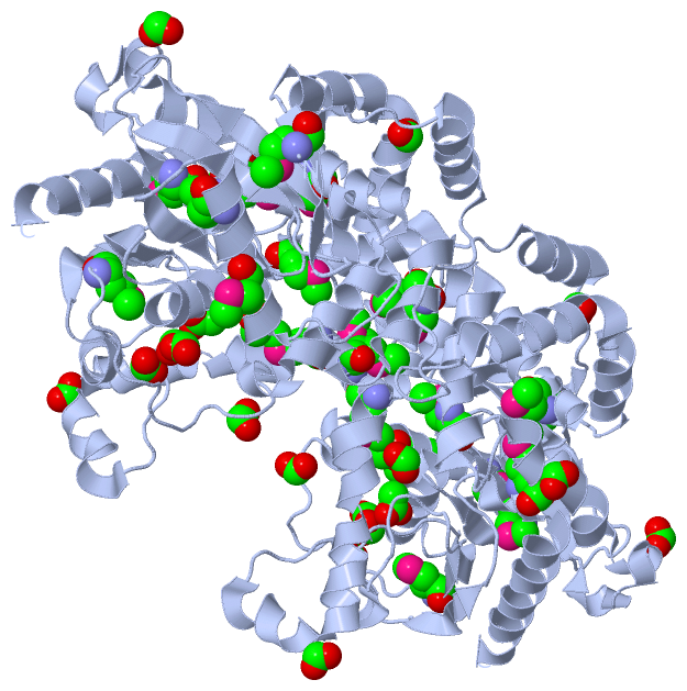
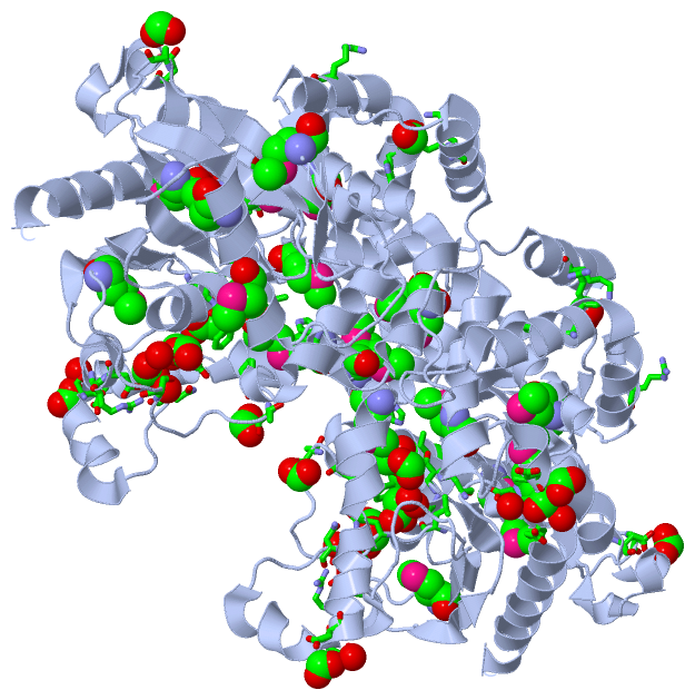
Still Images
|
||||||||||||||||
Databases
|
||||||||||||||||||||||||||||||||||||||||||||||||||||||||||||||||||||||||||||||||||||||||||||||||||||||||||||||||||||||||||||||||||||||||||||||||||||||||||||||||
Analysis Tools
|
|||||||||||||||||||||||||||||||||||||||||||||||||||||||||||||
Entries Sharing at Least One Protein Chain (UniProt ID)
Related Entries Specified in the PDB File
|
|