|
|
|
|
 Description
|
|
Compounds
|
||||||||||||||||||||||||||||||||||||||||||||||||
Chains, Units
Summary Information (see also Sequences/Alignments below) |
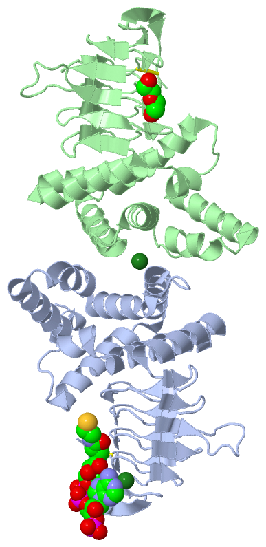
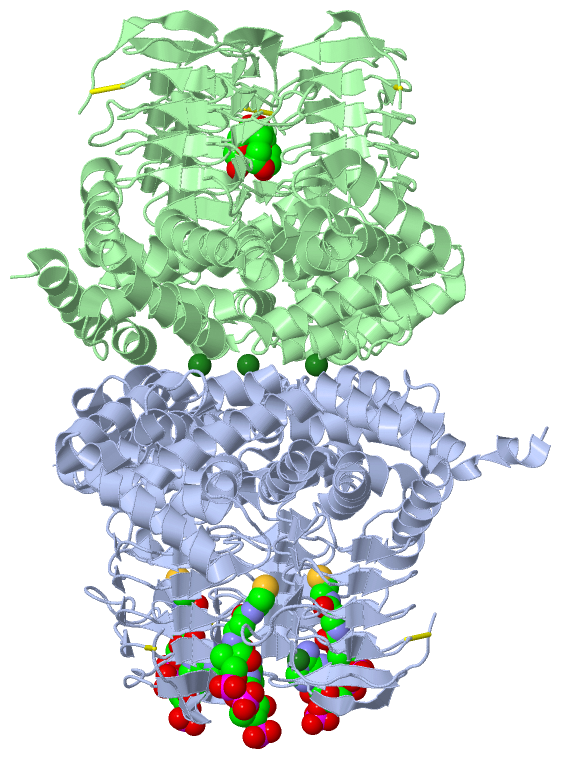
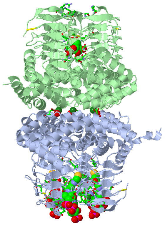
Ligands, Modified Residues, Ions (3, 4)| Asymmetric Unit (3, 4) Biological Unit 1 (2, 6) |
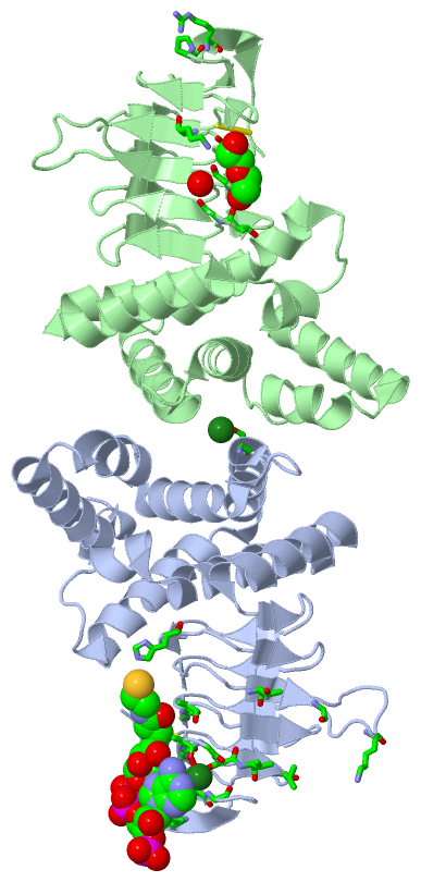
Sites (4, 4)
Asymmetric Unit (4, 4)
|
SS Bonds (2, 2)
Asymmetric Unit
|
||||||||||||
Cis Peptide Bonds (2, 2)
Asymmetric Unit
|
||||||||||||
SAPs(SNPs)/Variants (0, 0)| (no "SAP(SNP)/Variant" information available for 4HZD) |
PROSITE Motifs (0, 0)| (no "PROSITE Motif" information available for 4HZD) |
Exons (0, 0)| (no "Exon" information available for 4HZD) |
Sequences/Alignments
Asymmetric Unit
Chain A from PDB Type:PROTEIN Length:250
SCOP domains d4hzda_ A: automated matches SCOP domains
CATH domains ---------------------------------------------------------------------------------------------------------------------------------------------------------------------------------------------------------------------------------------------------------- CATH domains
Pfam domains ---------------------------------------------------------------------------------------------------------------------------------------------------------------------------------------------------------------------------------------------------------- Pfam domains
SAPs(SNPs) ---------------------------------------------------------------------------------------------------------------------------------------------------------------------------------------------------------------------------------------------------------- SAPs(SNPs)
PROSITE ---------------------------------------------------------------------------------------------------------------------------------------------------------------------------------------------------------------------------------------------------------- PROSITE
Transcript ---------------------------------------------------------------------------------------------------------------------------------------------------------------------------------------------------------------------------------------------------------- Transcript
4hzd A 11 AGLDQVDPIWHSIRAEAEEATRNDPVLGAFLYATILNQPSLEEAVMHRIAERLGHPDVSADILRQTFDTMLEANPEWSHVLRVDIQAVYDRDPAYSRFMDPVLYLKGFHAIQTHRLAHWLYKQGRKDFAYYLQSRSSSIFQTDIHPAARLGSGLFLDHATGLVVGETAVVEDNVSILHGVTLGGTGKSSGDRHPKIRQGVLIGAGAKILGNIQVGQCSKIAAGSVVLKSVPHNVTVAGVPARIIGETGCT 260
20 30 40 50 60 70 80 90 100 110 120 130 140 150 160 170 180 190 200 210 220 230 240 250 260
Chain B from PDB Type:PROTEIN Length:245
SCOP domains d4hzdb_ B: automated matches SCOP domains
CATH domains ----------------------------------------------------------------------------------------------------------------------------------------------------------------------------------------------------------------------------------------------------- CATH domains
Pfam domains ----------------------------------------------------------------------------------------------------------------------------------------------------------------------------------------------------------------------------------------------------- Pfam domains
SAPs(SNPs) ----------------------------------------------------------------------------------------------------------------------------------------------------------------------------------------------------------------------------------------------------- SAPs(SNPs)
PROSITE ----------------------------------------------------------------------------------------------------------------------------------------------------------------------------------------------------------------------------------------------------- PROSITE
Transcript ----------------------------------------------------------------------------------------------------------------------------------------------------------------------------------------------------------------------------------------------------- Transcript
4hzd B 16 VDPIWHSIRAEAEEATRNDPVLGAFLYATILNQPSLEEAVMHRIAERLGHPDVSADILRQTFDTMLEANPEWSHVLRVDIQAVYDRDPAYSRFMDPVLYLKGFHAIQTHRLAHWLYKQGRKDFAYYLQSRSSSIFQTDIHPAARLGSGLFLDHATGLVVGETAVVEDNVSILHGVTLGGTGKSSGDRHPKIRQGVLIGAGAKILGNIQVGQCSKIAAGSVVLKSVPHNVTVAGVPARIIGETGCT 260
25 35 45 55 65 75 85 95 105 115 125 135 145 155 165 175 185 195 205 215 225 235 245 255
|
||||||||||||||||||||
SCOP Domains (1, 2)
Asymmetric Unit
 |
CATH Domains (0, 0)| (no "CATH Domain" information available for 4HZD) |
Pfam Domains (0, 0)| (no "Pfam Domain" information available for 4HZD) |
Gene Ontology (0, 0)|
Asymmetric Unit(hide GO term definitions)
(no "Gene Ontology" information available for 4HZD)
|
Interactive Views
|
|||||||||||||||||||||||||||||||||||||||||||||||||||||||||||||||||||||||||||||||||||||||||||||||||||||||||||||||||||||||||||||||||||||||||||||||||||||||||||||||||||||||||||||||||||
Still Images
|
||||||||||||||||
Databases
|
||||||||||||||||||||||||||||||||||||||||||||||||||||||||||||||||||||||||||||||||||||||||||||||||||||||||||||||||||||||||||||||||||||||||||||||||||||||||||||||||
Analysis Tools
|
|||||||||||||||||||||||||||||||||||||||||||||||||||||||||||||
Entries Sharing at Least One Protein Chain (UniProt ID)
Related Entries Specified in the PDB File
|
|