|
|
|
|
 Description
|
|
Compounds
|
||||||||||||||||||||||||||||||||||||||||||||||||||||
Chains, Units
Summary Information (see also Sequences/Alignments below) |
Ligands, Modified Residues, Ions (1, 4)
Asymmetric Unit (1, 4)
|
Sites (4, 4)
Asymmetric Unit (4, 4)
|
SS Bonds (0, 0)| (no "SS Bond" information available for 3P1B) |
Cis Peptide Bonds (0, 0)| (no "Cis Peptide Bond" information available for 3P1B) |
SAPs(SNPs)/Variants (0, 0)| (no "SAP(SNP)/Variant" information available for 3P1B) |
PROSITE Motifs (0, 0)| (no "PROSITE Motif" information available for 3P1B) |
Exons (0, 0)| (no "Exon" information available for 3P1B) |
Sequences/Alignments
Asymmetric UnitChain A from PDB Type:PROTEIN Length:266 aligned with Q9U8X2_ENTHI | Q9U8X2 from UniProtKB/TrEMBL Length:305 Alignment length:269 1 | 9 19 29 39 49 59 69 79 89 99 109 119 129 139 149 159 169 179 189 199 209 219 229 239 249 259 Q9U8X2_ENTHI - -MDNYIYSIAHQLYEMYLQDEDAFHSKRDYPHKKVFTELQKLRKIFFPDFFMKHQKITESHIASELTKLVDYIKDSVTAYNDELFAHQCVMAILEKLPSIKRTLKTDLIAAYAGDPAAPGLSLIIRCYPGFQAVIVYRIAHVLYECGERYYCREMMESVHSYTSIDIHPGASIKGHFFIDHGVGVVIGETAIIGEWCRIYQSVTLGAMHFQEEGGVIKRGTKRHPTVGDYVTIGTGAKVLGNIIVGSHVRIGANCWIDRDVDSNQTVYI 268 SCOP domains ----------------------------------------------------------------------------------------------------------------------------------------------------------------------------------------------------------------------------------------------------------------------------- SCOP domains CATH domains ----------------------------------------------------------------------------------------------------------------------------------------------------------------------------------------------------------------------------------------------------------------------------- CATH domains Pfam domains ---------------------------------------------------SATase_N-3p1bA02 A:51-135 ---------------------------------------------------------------------------------------Hexapep-3p1bA01 A:223-258 ---------- Pfam domains SAPs(SNPs) ----------------------------------------------------------------------------------------------------------------------------------------------------------------------------------------------------------------------------------------------------------------------------- SAPs(SNPs) PROSITE ----------------------------------------------------------------------------------------------------------------------------------------------------------------------------------------------------------------------------------------------------------------------------- PROSITE Transcript ----------------------------------------------------------------------------------------------------------------------------------------------------------------------------------------------------------------------------------------------------------------------------- Transcript 3p1b A 0 AMDNYIYSIAHQLYEMYLQDEDAFHSKRDYPHKKVFTELQKLRKIFFPDFFMKHQKITESHIASELTKLVDYIKDSVTAYNDELFAHQCVMAILEKLPSIKRTLKTDLIAAYAGDPAAPGLSLIIRCYPGFQAVIVYRIAHVLYECGERYYCREMMESVHSYTSIDIHPGASIKGHFFIDHGVGVVIGETAIIGEWCRIYQSVTLGAMHFQE---VIKRGTKRHPTVGDYVTIGTGAKVLGNIIVGSHVRIGANCWIDRDVDSNQTVYI 268 9 19 29 39 49 59 69 79 89 99 109 119 129 139 149 159 169 179 189 199 209 | | 219 229 239 249 259 211 215
|
||||||||||||||||||||
SCOP Domains (0, 0)| (no "SCOP Domain" information available for 3P1B) |
CATH Domains (0, 0)| (no "CATH Domain" information available for 3P1B) |
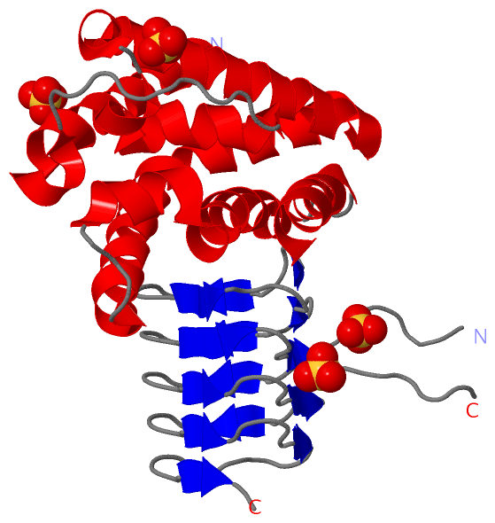
Pfam Domains (2, 2)| Asymmetric Unit |
Gene Ontology (1, 1)|
Asymmetric Unit(hide GO term definitions) Chain A (Q9U8X2_ENTHI | Q9U8X2)
|
||||||||||||
Interactive Views
|
|||||||||||||||||||||||||||||||||||||||||||||||||||||||||||||||||||||||||||||||||||||||||||||||||||||||||||||||||||||||||||||||||||||||||||||||||||||||||||||
Still Images
|
||||||||||||||||
Databases
|
||||||||||||||||||||||||||||||||||||||||||||||||||||||||||||||||||||||||||||||||||||||||||||||||||||||||||||||||||||||||||||||||||||||||||||||||||||||||||||||||
Analysis Tools
|
|||||||||||||||||||||||||||||||||||||||||||||||||||||||||||||
Entries Sharing at Least One Protein Chain (UniProt ID)
Related Entries Specified in the PDB File
|
|