Show PDB file:   
         Plain Text   HTML   (compressed file size)
QuickSearch:   
by PDB,NDB,UniProt,PROSITE Code or Search Term(s)  
(-)Asymmetric Unit
(-)Asym. Unit - sites
(-)Biological Unit 1
(-)Biol. Unit 1 - sites
collapse expand < >
Image Asymmetric Unit
Asymmetric Unit  (Jmol Viewer)
Image Asym. Unit - sites
Asym. Unit - sites  (Jmol Viewer)
Image Biological Unit 1
Biological Unit 1  (Jmol Viewer)
Image Biol. Unit 1 - sites
Biol. Unit 1 - sites  (Jmol Viewer)

(-) Description

Title :  CRYSTAL STRUCTURE OF A PARALLEL 4-HELIX COILED COIL CC-HEX-II- 22
 
Authors :  B. Chi, N. R. Zaccai, R. L. Brady, D. N. Woolfson
Date :  22 Sep 12  (Deposition) - 05 Feb 14  (Release) - 05 Feb 14  (Revision)
Method :  X-RAY DIFFRACTION
Resolution :  1.88
Chains :  Asym. Unit :  A,B
Biol. Unit 1:  A,B  (2x)
Keywords :  Cc-Hex Related, Synthetic Coiled Coil Peptide, N-Terminal Acetylation; C-Terminal Amidation, De Novo Protein (Keyword Search: [Gene Ontology, PubMed, Web (Google))
 
Reference :  B. Chi, N. R. Zaccai, R. L. Brady, D. N. Woolfson
Tba
J. Am. Chem. Soc.
PubMed: search

(-) Compounds

Molecule 1 - CC-HEX-II-PHI22
    ChainsA, B
    EngineeredYES
    Organism ScientificSYNTHETIC CONSTRUCT
    Organism Taxid32630
    Other DetailsSTANDARD F-MOC PEPTIDE SYNTHESIS
    SyntheticYES

 Structural Features

(-) Chains, Units

  12
Asymmetric Unit AB
Biological Unit 1 (2x)AB

Summary Information (see also Sequences/Alignments below)

(-) Ligands, Modified Residues, Ions  (5, 10)

Asymmetric Unit (5, 10)
No.NameCountTypeFull Name
1ACE2Mod. Amino AcidACETYL GROUP
2GOL1Ligand/IonGLYCEROL
3NH22Mod. Amino AcidAMINO GROUP
4PHI2Mod. Amino AcidIODO-PHENYLALANINE
5TBU3Ligand/IonTERTIARY-BUTYL ALCOHOL
Biological Unit 1 (5, 20)
No.NameCountTypeFull Name
1ACE4Mod. Amino AcidACETYL GROUP
2GOL2Ligand/IonGLYCEROL
3NH24Mod. Amino AcidAMINO GROUP
4PHI4Mod. Amino AcidIODO-PHENYLALANINE
5TBU6Ligand/IonTERTIARY-BUTYL ALCOHOL

(-) Sites  (4, 4)

Asymmetric Unit (4, 4)
No.NameEvidenceResiduesDescription
1AC1SOFTWAREALA A:28 , ALA B:28BINDING SITE FOR RESIDUE TBU A 101
2AC2SOFTWAREGLU A:16 , GLU A:23 , HOH A:218 , HOH B:216BINDING SITE FOR RESIDUE GOL A 102
3AC3SOFTWAREILE A:3 , ALA A:7 , ILE B:3 , ALA B:7 , ILE B:10BINDING SITE FOR RESIDUE TBU A 103
4AC4SOFTWAREILE A:17 , ALA A:21 , ALA B:21 , ILE B:24BINDING SITE FOR RESIDUE TBU B 101

(-) SS Bonds  (0, 0)

(no "SS Bond" information available for 4H8F)

(-) Cis Peptide Bonds  (1, 1)

Asymmetric Unit
No.Residues
1Gly A:1 -Glu A:2

 Sequence-Structure Mapping

(-) SAPs(SNPs)/Variants  (0, 0)

(no "SAP(SNP)/Variant" information available for 4H8F)

(-) PROSITE Motifs  (0, 0)

(no "PROSITE Motif" information available for 4H8F)

(-) Exons   (0, 0)

(no "Exon" information available for 4H8F)

(-) Sequences/Alignments

Asymmetric Unit
   Reformat: Number of residues per line =  ('0' or empty: single-line sequence representation)
  Number of residues per labelling interval =   
  UniProt sequence: complete  aligned part    
   Show mapping: SCOP domains CATH domains Pfam domains Secondary structure (by author)
SAPs(SNPs) PROSITE motifs Exons
(details for a mapped element are shown in a popup box when the mouse pointer rests over it)
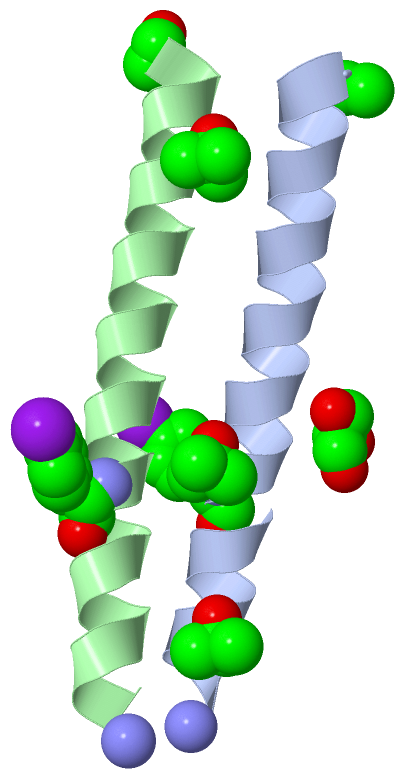
Chain A from PDB  Type:PROTEIN  Length:33
                                                                
               SCOP domains --------------------------------- SCOP domains
               CATH domains --------------------------------- CATH domains
               Pfam domains --------------------------------- Pfam domains
         Sec.struct. author ..hhhhhhhhhhhhhhhhhhhhhhhhhhhhhh. Sec.struct. author
                 SAPs(SNPs) --------------------------------- SAPs(SNPs)
                    PROSITE --------------------------------- PROSITE
                 Transcript --------------------------------- Transcript
                  4h8f A  0 xGEIKAIAQEIKAIAKEIKAIAfEIKAIAQGYx 32
                            |        9        19  |     29  |
                            |                    22-PHI    32-NH2
                            0-ACE                            

Chain B from PDB  Type:PROTEIN  Length:33
                                                                
               SCOP domains --------------------------------- SCOP domains
               CATH domains --------------------------------- CATH domains
               Pfam domains --------------------------------- Pfam domains
         Sec.struct. author .hhhhhhhhhhhhhhhhhhhhhhhhhhhhhhh. Sec.struct. author
                 SAPs(SNPs) --------------------------------- SAPs(SNPs)
                    PROSITE --------------------------------- PROSITE
                 Transcript --------------------------------- Transcript
                  4h8f B  0 xGEIKAIAQEIKAIAKEIKAIAfEIKAIAQGYx 32
                            |        9        19  |     29  |
                            |                    22-PHI    32-NH2
                            0-ACE                            

   Legend:   → Mismatch (orange background)
  - → Gap (green background, '-', border residues have a numbering label)
    → Modified Residue (blue background, lower-case, 'x' indicates undefined single-letter code, labelled with number + name)
  x → Chemical Group (purple background, 'x', labelled with number + name, e.g. ACE or NH2)
  extra numbering lines below/above indicate numbering irregularities and modified residue names etc., number ends below/above '|'

 Classification and Annotation

(-) SCOP Domains  (0, 0)

(no "SCOP Domain" information available for 4H8F)

(-) CATH Domains  (0, 0)

(no "CATH Domain" information available for 4H8F)

(-) Pfam Domains  (0, 0)

(no "Pfam Domain" information available for 4H8F)

(-) Gene Ontology  (0, 0)

Asymmetric Unit(hide GO term definitions)
    (no "Gene Ontology" information available for 4H8F)

 Visualization

(-) Interactive Views

Asymmetric Unit
  Complete Structure
    Jena3D(integrated viewing of ligand, site, SAP, PROSITE, SCOP information)
    WebMol | AstexViewer[tm]@PDBe
(Java Applets, require no local installation except for Java; loading may be slow)
    STRAP
(Java WebStart application, automatic local installation, requires Java; full application with system access!)
    RasMol
(require local installation)
    Molscript (VRML)
(requires installation of a VRML viewer; select preferred view via VRML and generate a mono or stereo PDF format file)
 
  Ligands, Modified Residues, Ions
    ACE  [ RasMol | Jena3D ]  +environment [ RasMol | Jena3D ]
    GOL  [ RasMol | Jena3D ]  +environment [ RasMol | Jena3D ]
    NH2  [ RasMol | Jena3D ]  +environment [ RasMol | Jena3D ]
    PHI  [ RasMol | Jena3D ]  +environment [ RasMol | Jena3D ]
    TBU  [ RasMol | Jena3D ]  +environment [ RasMol | Jena3D ]
 
  Sites
    AC1  [ RasMol ]  +environment [ RasMol ]
    AC2  [ RasMol ]  +environment [ RasMol ]
    AC3  [ RasMol ]  +environment [ RasMol ]
    AC4  [ RasMol ]  +environment [ RasMol ]
 
  Cis Peptide Bonds
    Gly A:1 - Glu A:2   [ RasMol ]  
 
Biological Unit
  Complete Structure
    Biological Unit 1  [ Jena3D ]

(-) Still Images

Jmol
  protein: cartoon or spacefill or dots and stick; nucleic acid: cartoon and stick; ligands: spacefill; active site: stick
Molscript
  protein, nucleic acid: cartoon; ligands: spacefill; active site: ball and stick

 Databases and Analysis Tools

(-) Databases

Access by PDB/NDB ID
  4h8f
    Family and Domain InformationProDom | SYSTERS
    General Structural InformationGlycoscienceDB | MMDB | NDB | OCA | PDB | PDBe | PDBj | PDBsum | PDBWiki | PQS | PROTEOPEDIA
    Orientation in MembranesOPM
    Protein SurfaceSURFACE
    Secondary StructureDSSP (structure derived) | HSSP (homology derived)
    Structural GenomicsGeneCensus
    Structural NeighboursCE | VAST
    Structure ClassificationCATH | Dali | SCOP
    Validation and Original DataBMRB Data View | BMRB Restraints Grid | EDS | PROCHECK | RECOORD | WHAT_CHECK
 
Access by UniProt ID/Accession number
  (no 'UniProt ID/Accession number' available) |
    Comparative Protein Structure ModelsModBase
    Genomic InformationEnsembl
    Protein-protein InteractionDIP
    Sequence, Family and Domain InformationInterPro | Pfam | SMART | UniProt
 
Access by Enzyme Classificator   (EC Number)
  (no 'Enzyme Classificator' available)
    General Enzyme InformationBRENDA | EC-PDB | Enzyme | IntEnz
    PathwayKEGG | MetaCyc
 
Access by Disease Identifier   (MIM ID)
  (no 'MIM ID' available)
    Disease InformationOMIM
 
Access by GenAge ID
  (no 'GenAge ID' available)
    Age Related InformationGenAge

(-) Analysis Tools

Access by PDB/NDB ID
    Domain InformationXDom
    Interatomic Contacts of Structural UnitsCSU
    Ligand-protein ContactsLPC
    Protein CavitiescastP
    Sequence and Secondary StructurePDBCartoon
    Structure AlignmentSTRAP(Java WebStart application, automatic local installation, requires Java; full application with system access!)
    Structure and Sequence BrowserSTING
 
Access by UniProt ID/Accession number
  (no 'UniProt ID/Accession number' available) |
    Protein Disorder PredictionDisEMBL | FoldIndex | GLOBPLOT

 Related Entries

(-) Entries Sharing at Least One Protein Chain (UniProt ID)

(no "Entries Sharing at Least One Protein Chain" available for 4H8F)

(-) Related Entries Specified in the PDB File

3r3k
3r4a
3r4h
4h7r POSITION 22 W MUTANT
4h8g
4h8l
4h8m
4h8o