|
|
|
|
 Description
|
|
Compounds
|
||||||||||||||||||||||||||||||||||||||||||||||||||||||||
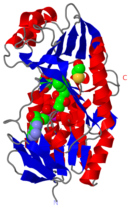
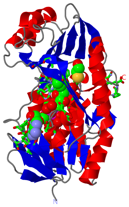
Chains, Units
Summary Information (see also Sequences/Alignments below) |
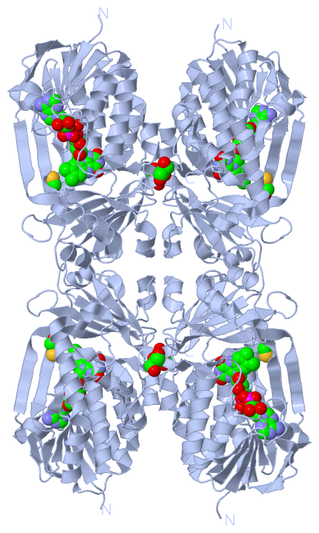
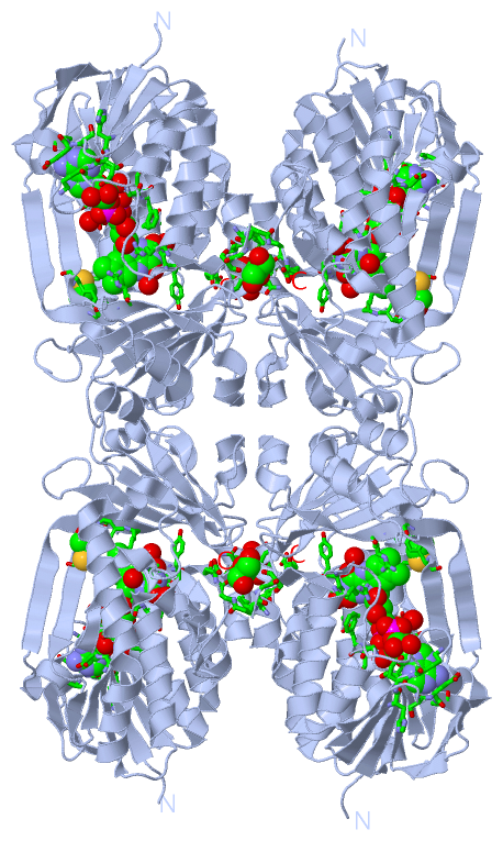
Ligands, Modified Residues, Ions (3, 3)| Asymmetric Unit (3, 3) Biological Unit 1 (3, 12) |
Sites (3, 3)
Asymmetric Unit (3, 3)
|
SS Bonds (0, 0)| (no "SS Bond" information available for 4H2N) |
Cis Peptide Bonds (2, 2)
Asymmetric Unit
|
||||||||||||
SAPs(SNPs)/Variants (0, 0)| (no "SAP(SNP)/Variant" information available for 4H2N) |
PROSITE Motifs (0, 0)| (no "PROSITE Motif" information available for 4H2N) |
Exons (0, 0)| (no "Exon" information available for 4H2N) |
Sequences/Alignments
Asymmetric Unit
Chain A from PDB Type:PROTEIN Length:370
SCOP domains ---------------------------------------------------------------------------------------------------------------------------------------------------------------------------------------------------------------------------------------------------------------------------------------------------------------------------------------------------------------------------------- SCOP domains
CATH domains ---------------------------------------------------------------------------------------------------------------------------------------------------------------------------------------------------------------------------------------------------------------------------------------------------------------------------------------------------------------------------------- CATH domains
Pfam domains ---------------------------------------------------------------------------------------------------------------------------------------------------------------------------------------------------------------------------------------------------------------------------------------------------------------------------------------------------------------------------------- Pfam domains
SAPs(SNPs) ---------------------------------------------------------------------------------------------------------------------------------------------------------------------------------------------------------------------------------------------------------------------------------------------------------------------------------------------------------------------------------- SAPs(SNPs)
PROSITE ---------------------------------------------------------------------------------------------------------------------------------------------------------------------------------------------------------------------------------------------------------------------------------------------------------------------------------------------------------------------------------- PROSITE
Transcript ---------------------------------------------------------------------------------------------------------------------------------------------------------------------------------------------------------------------------------------------------------------------------------------------------------------------------------------------------------------------------------- Transcript
4h2n A 10 KTRRAEVAGGGFAGLTAAIALKQNGWDVRLHEKSSELRAFGAGIYLWHNGLRVLEGLGALDDVLQGSHTPPTYETWMHNKSVSKETFNGLPWRIMTRSHLHDALVNRARALGVDISVNSEAVAADPVGRLTLQTGEVLEADLIVGADGVGSKVRDSIGFKQDRWVSKDGLIRLIVPRMKKELGHGEWDNTIDMWNFWPRVQRILYSPCNENELYLGLMAPAADPRGSSVPIDLEVWVEMFPFLEPCLIEAAKLKTARYDKFETTKLDSWTRGKVALVGDAAHAMCPALAQGAGCAMVNAFSLSQDLEEGSSVEDALVAWETRIRPITDRCQALSGDYAANRSLSKGNMFTPAALEAARYDPLRRVYSWPQ 379
19 29 39 49 59 69 79 89 99 109 119 129 139 149 159 169 179 189 199 209 219 229 239 249 259 269 279 289 299 309 319 329 339 349 359 369 379
|
||||||||||||||||||||
SCOP Domains (0, 0)| (no "SCOP Domain" information available for 4H2N) |
CATH Domains (0, 0)| (no "CATH Domain" information available for 4H2N) |
Pfam Domains (0, 0)| (no "Pfam Domain" information available for 4H2N) |
Gene Ontology (4, 4)|
Asymmetric Unit(hide GO term definitions) |
Interactive Views
|
||||||||||||||||||||||||||||||||||||||||||||||||||||||||||||||||||||||||||||||||||||||||||||||||||||||||||||||||||||||||||||||||||||||||||||||||||||||||||||||||||||||||||||
Still Images
|
||||||||||||||||
Databases
|
||||||||||||||||||||||||||||||||||||||||||||||||||||||||||||||||||||||||||||||||||||||||||||||||||||||||||||||||||||||||||||||||||||||||||||||||||||||||||||||||
Analysis Tools
|
|||||||||||||||||||||||||||||||||||||||||||||||||||||||||||||
Entries Sharing at Least One Protein Chain (UniProt ID)
Related Entries Specified in the PDB File
|
|