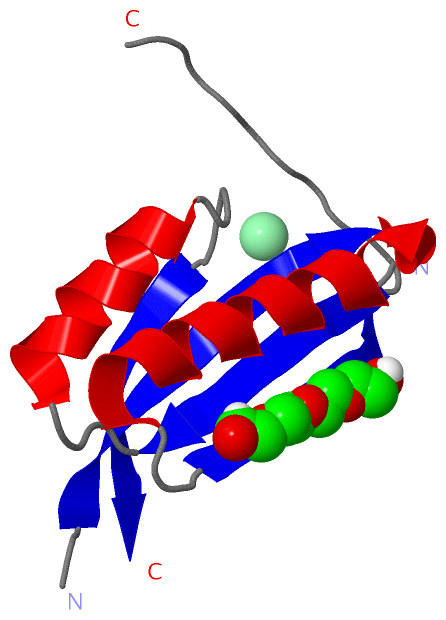
Show PDB file:   
         Plain Text   HTML   (compressed file size)
QuickSearch:   
by PDB,NDB,UniProt,PROSITE Code or Search Term(s)  
(-)Asymmetric Unit
(-)Asym. Unit - sites
(-)Biological Unit 1
(-)Biol. Unit 1 - sites
collapse expand < >
Image Asymmetric Unit
Asymmetric Unit  (Jmol Viewer)
Image Asym. Unit - sites
Asym. Unit - sites  (Jmol Viewer)
Image Biological Unit 1
Biological Unit 1  (Jmol Viewer)
Image Biol. Unit 1 - sites
Biol. Unit 1 - sites  (Jmol Viewer)

(-) Description

Title :  STRUCTURE OF WILDTYPE PII FROM S. ELONGATUS AT HIGH RESOLUTION
 
Authors :  K. Zeth, K. Forchhammer
Date :  25 Aug 13  (Deposition) - 15 Jan 14  (Release) - 03 Dec 14  (Revision)
Method :  X-RAY DIFFRACTION
Resolution :  1.60
Chains :  Asym. Unit :  A
Biol. Unit 1:  A  (3x)
Keywords :  Transcription (Keyword Search: [Gene Ontology, PubMed, Web (Google))
 
Reference :  K. Zeth, O. Fokina, K. Forchhammer
Structural Basis And Target-Specific Modulation Of Adp Sensing By The Synechococcus Elongatus Pii Signaling Protein.
J. Biol. Chem. V. 289 8960 2014
PubMed-ID: 24519945  |  Reference-DOI: 10.1074/JBC.M113.536557

(-) Compounds

Molecule 1 - NITROGEN REGULATORY PROTEIN P-II
    ChainsA
    EngineeredYES
    Expression SystemESCHERICHIA COLI
    Expression System StrainBL21(DE3)
    Expression System Taxid469008
    Expression System VectorPET28
    Expression System Vector TypePLASMID
    Organism ScientificSYNECHOCOCCUS ELONGATUS
    Organism Taxid32046
    SynonymPII SIGNAL TRANSDUCING PROTEIN, PII

 Structural Features

(-) Chains, Units

  1
Asymmetric Unit A
Biological Unit 1 (3x)A

Summary Information (see also Sequences/Alignments below)

(-) Ligands, Modified Residues, Ions  (2, 2)

Asymmetric Unit (2, 2)
No.NameCountTypeFull Name
1CL1Ligand/IonCHLORIDE ION
2PG41Ligand/IonTETRAETHYLENE GLYCOL
Biological Unit 1 (1, 3)
No.NameCountTypeFull Name
1CL-1Ligand/IonCHLORIDE ION
2PG43Ligand/IonTETRAETHYLENE GLYCOL

(-) Sites  (2, 2)

Asymmetric Unit (2, 2)
No.NameEvidenceResiduesDescription
1AC1SOFTWAREVAL A:64 , LYS A:90 , PHE A:92 , ARG A:101BINDING SITE FOR RESIDUE CL A1107
2AC2SOFTWAREASP A:71 , ARG A:82 , GLN A:98BINDING SITE FOR RESIDUE PG4 A1108

(-) SS Bonds  (0, 0)

(no "SS Bond" information available for 4C3L)

(-) Cis Peptide Bonds  (1, 1)

Asymmetric Unit
No.Residues
1Glu A:85 -Ile A:86

 Sequence-Structure Mapping

(-) SAPs(SNPs)/Variants  (0, 0)

(no "SAP(SNP)/Variant" information available for 4C3L)

(-) PROSITE Motifs  (2, 2)

Asymmetric Unit (2, 2)
 PROSITEUniProtKBPDB
No.IDACDescriptionIDLocationCountLocation
1PII_GLNB_DOMPS51343 P-II protein family profile.GLNB_SYNE71-112  1A:1-106
2PII_GLNB_CTERPS00638 P-II protein C-terminal region signature.GLNB_SYNE783-96  1A:83-96
Biological Unit 1 (2, 6)
 PROSITEUniProtKBPDB
No.IDACDescriptionIDLocationCountLocation
1PII_GLNB_DOMPS51343 P-II protein family profile.GLNB_SYNE71-112  3A:1-106
2PII_GLNB_CTERPS00638 P-II protein C-terminal region signature.GLNB_SYNE783-96  3A:83-96

(-) Exons   (0, 0)

(no "Exon" information available for 4C3L)

(-) Sequences/Alignments

Asymmetric Unit
   Reformat: Number of residues per line =  ('0' or empty: single-line sequence representation)
  Number of residues per labelling interval =   
  UniProt sequence: complete  aligned part    
   Show mapping: SCOP domains CATH domains Pfam domains Secondary structure (by author)
SAPs(SNPs) PROSITE motifs Exons
(details for a mapped element are shown in a popup box when the mouse pointer rests over it)
Chain A from PDB  Type:PROTEIN  Length:89
 aligned with GLNB_SYNE7 | P0A3F4 from UniProtKB/Swiss-Prot  Length:112

    Alignment length:106
                                    10        20        30        40        50        60        70        80        90       100      
           GLNB_SYNE7     1 MKKIEAIIRPFKLDEVKIALVNAGIVGMTVSEVRGFGRQKGQTERYRGSEYTVEFLQKLKLEIVVEDAQVDTVIDKIVAAARTGEIGDGKIFVSPVDQTIRIRTGE 106
               SCOP domains d4c3la_ A: PII (product of glnB)                                                                           SCOP domains
               CATH domains ---------------------------------------------------------------------------------------------------------- CATH domains
               Pfam domains ---------------------------------------------------------------------------------------------------------- Pfam domains
         Sec.struct. author .eeeeeee...hhhhhhhhhhhh.....eeeeeee.-----------------..eeeeeeeeeehhhhhhhhhhhhhhhhhhhh...eeeeeee........... Sec.struct. author
                 SAPs(SNPs) ---------------------------------------------------------------------------------------------------------- SAPs(SNPs)
                PROSITE (1) PII_GLNB_DOM  PDB: A:1-106 UniProt: 1-112                                                                  PROSITE (1)
                PROSITE (2) ----------------------------------------------------------------------------------PII_GLNB_CTER ---------- PROSITE (2)
                 Transcript ---------------------------------------------------------------------------------------------------------- Transcript
                 4c3l A   1 MKKIEAIIRPFKLDEVKIALVNAGIVGMTVSEVRGF-----------------EFLQKLKLEIVVEDAQVDTVIDKIVAAARTGEIGDGKIFVSPVDQTIRIRTGE 106
                                    10        20        30     |   -         -   |    60        70        80        90       100      
                                                              36                54                                                    

   Legend:   → Mismatch (orange background)
  - → Gap (green background, '-', border residues have a numbering label)
    → Modified Residue (blue background, lower-case, 'x' indicates undefined single-letter code, labelled with number + name)
  x → Chemical Group (purple background, 'x', labelled with number + name, e.g. ACE or NH2)
  extra numbering lines below/above indicate numbering irregularities and modified residue names etc., number ends below/above '|'

 Classification and Annotation

(-) SCOP Domains  (1, 1)

Asymmetric Unit

(-) CATH Domains  (0, 0)

(no "CATH Domain" information available for 4C3L)

(-) Pfam Domains  (0, 0)

(no "Pfam Domain" information available for 4C3L)

(-) Gene Ontology  (8, 8)

Asymmetric Unit(hide GO term definitions)
Chain A   (GLNB_SYNE7 | P0A3F4)
molecular function
    GO:0030234    enzyme regulator activity    Binds to and modulates the activity of an enzyme.
    GO:0042802    identical protein binding    Interacting selectively and non-covalently with an identical protein or proteins.
    GO:0000166    nucleotide binding    Interacting selectively and non-covalently with a nucleotide, any compound consisting of a nucleoside that is esterified with (ortho)phosphate or an oligophosphate at any hydroxyl group on the ribose or deoxyribose.
    GO:0005515    protein binding    Interacting selectively and non-covalently with any protein or protein complex (a complex of two or more proteins that may include other nonprotein molecules).
biological process
    GO:0050790    regulation of catalytic activity    Any process that modulates the activity of an enzyme.
    GO:0006808    regulation of nitrogen utilization    Any process that modulates the frequency, rate or extent of nitrogen utilization.
    GO:0006355    regulation of transcription, DNA-templated    Any process that modulates the frequency, rate or extent of cellular DNA-templated transcription.
    GO:0006351    transcription, DNA-templated    The cellular synthesis of RNA on a template of DNA.

 Visualization

(-) Interactive Views

Asymmetric Unit
  Complete Structure
    Jena3D(integrated viewing of ligand, site, SAP, PROSITE, SCOP information)
    WebMol | AstexViewer[tm]@PDBe
(Java Applets, require no local installation except for Java; loading may be slow)
    STRAP
(Java WebStart application, automatic local installation, requires Java; full application with system access!)
    RasMol
(require local installation)
    Molscript (VRML)
(requires installation of a VRML viewer; select preferred view via VRML and generate a mono or stereo PDF format file)
 
  Ligands, Modified Residues, Ions
    CL  [ RasMol | Jena3D ]  +environment [ RasMol | Jena3D ]
    PG4  [ RasMol | Jena3D ]  +environment [ RasMol | Jena3D ]
 
  Sites
    AC1  [ RasMol ]  +environment [ RasMol ]
    AC2  [ RasMol ]  +environment [ RasMol ]
 
  Cis Peptide Bonds
    Glu A:85 - Ile A:86   [ RasMol ]  
 
Biological Unit
  Complete Structure
    Biological Unit 1  [ Jena3D ]

(-) Still Images

Jmol
  protein: cartoon or spacefill or dots and stick; nucleic acid: cartoon and stick; ligands: spacefill; active site: stick
Molscript
  protein, nucleic acid: cartoon; ligands: spacefill; active site: ball and stick

 Databases and Analysis Tools

(-) Databases

Access by PDB/NDB ID
  4c3l
    Family and Domain InformationProDom | SYSTERS
    General Structural InformationGlycoscienceDB | MMDB | NDB | OCA | PDB | PDBe | PDBj | PDBsum | PDBWiki | PQS | PROTEOPEDIA
    Orientation in MembranesOPM
    Protein SurfaceSURFACE
    Secondary StructureDSSP (structure derived) | HSSP (homology derived)
    Structural GenomicsGeneCensus
    Structural NeighboursCE | VAST
    Structure ClassificationCATH | Dali | SCOP
    Validation and Original DataBMRB Data View | BMRB Restraints Grid | EDS | PROCHECK | RECOORD | WHAT_CHECK
 
Access by UniProt ID/Accession number
  GLNB_SYNE7 | P0A3F4
    Comparative Protein Structure ModelsModBase
    Genomic InformationEnsembl
    Protein-protein InteractionDIP
    Sequence, Family and Domain InformationInterPro | Pfam | SMART | UniProtKB/SwissProt
 
Access by Enzyme Classificator   (EC Number)
  (no 'Enzyme Classificator' available)
    General Enzyme InformationBRENDA | EC-PDB | Enzyme | IntEnz
    PathwayKEGG | MetaCyc
 
Access by Disease Identifier   (MIM ID)
  (no 'MIM ID' available)
    Disease InformationOMIM
 
Access by GenAge ID
  (no 'GenAge ID' available)
    Age Related InformationGenAge

(-) Analysis Tools

Access by PDB/NDB ID
    Domain InformationXDom
    Interatomic Contacts of Structural UnitsCSU
    Ligand-protein ContactsLPC
    Protein CavitiescastP
    Sequence and Secondary StructurePDBCartoon
    Structure AlignmentSTRAP(Java WebStart application, automatic local installation, requires Java; full application with system access!)
    Structure and Sequence BrowserSTING
 
Access by UniProt ID/Accession number
  GLNB_SYNE7 | P0A3F4
    Protein Disorder PredictionDisEMBL | FoldIndex | GLOBPLOT (for more information see DisProt)

 Related Entries

(-) Entries Sharing at Least One Protein Chain (UniProt ID)

UniProtKB/Swiss-Prot
        GLNB_SYNE7 | P0A3F41qy7 2jj4 2v5h 2xbp 2xg8 2xul 2xzw 4aff 4c3k 4c3m

(-) Related Entries Specified in the PDB File

4c3k STRUCTURE OF MIXED PII-ADP COMPLEXES FROM S. ELONGATUS
4c3m STRUCTURE OF WILDTYPE PII FROM S. ELONGATUS AT MEDIUM RESOLUTION