Show PDB file:   
         Plain Text   HTML   (compressed file size)
QuickSearch:   
by PDB,NDB,UniProt,PROSITE Code or Search Term(s)  
(-)Asymmetric Unit
(-)Asym. Unit - sites
(-)Biological Unit 1
(-)Biol. Unit 1 - sites
collapse expand < >
Image Asymmetric Unit
Asymmetric Unit  (Jmol Viewer)
Image Asym. Unit - sites
Asym. Unit - sites  (Jmol Viewer)
Image Biological Unit 1
Biological Unit 1  (Jmol Viewer)
Image Biol. Unit 1 - sites
Biol. Unit 1 - sites  (Jmol Viewer)

(-) Description

Title :  STRUCTURE OF THE HUMAN EOSINOPHIL CATIONIC PROTEIN IN COMPLEX WITH SULFATE ANIONS
 
Authors :  E. Boix, D. Pulido, M. Moussaoui, V. Nogues, S. Russi
Date :  28 Sep 11  (Deposition) - 27 Jun 12  (Release) - 27 Jun 12  (Revision)
Method :  X-RAY DIFFRACTION
Resolution :  1.69
Chains :  Asym. Unit :  A,B
Biol. Unit 1:  A,B  (2x)
Keywords :  Hydrolase, Oxidoreductase, Antimicrobial, Cytotoxic (Keyword Search: [Gene Ontology, PubMed, Web (Google))
 
Reference :  E. Boix, D. Pulido, M. Moussaoui, V. Nogues, S. Russi
The Sulfate-Binding Site Structure Of The Human Eosinophil Cationic Protein As Revealed By A New Crystal Form.
J. Struct. Biol. V. 179 1 2012
PubMed-ID: 22579681  |  Reference-DOI: 10.1016/J.JSB.2012.04.023

(-) Compounds

Molecule 1 - EOSINOPHIL CATIONIC PROTEIN
    CellEOSINOPHIL
    ChainsA, B
    EC Number3.1.27.-
    EngineeredYES
    Expression SystemESCHERICHIA COLI
    Expression System PlasmidPET11C/ECP
    Expression System StrainBL21(DE3)
    Expression System Taxid469008
    Expression System VectorPET11C
    Expression System Vector TypePLASMID
    Organism CommonHUMAN
    Organism ScientificHOMO SAPIENS
    Organism Taxid9606
    SynonymECP, RIBONUCLEASE 3, RNASE 3

 Structural Features

(-) Chains, Units

  12
Asymmetric Unit AB
Biological Unit 1 (2x)AB

Summary Information (see also Sequences/Alignments below)

(-) Ligands, Modified Residues, Ions  (1, 14)

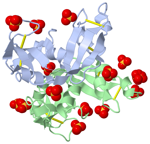
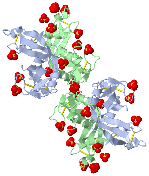
Asymmetric Unit (1, 14)
No.NameCountTypeFull Name
1SO414Ligand/IonSULFATE ION
Biological Unit 1 (1, 28)
No.NameCountTypeFull Name
1SO428Ligand/IonSULFATE ION

(-) Sites  (14, 14)

Asymmetric Unit (14, 14)
No.NameEvidenceResiduesDescription
01AC1SOFTWAREARG A:34 , ARG A:36 , LYS A:38 , ASN A:39 , HOH A:2074 , HOH A:2075 , ARG B:104BINDING SITE FOR RESIDUE SO4 A1134
02AC2SOFTWAREARG A:1 , ARG A:7 , HOH A:2191BINDING SITE FOR RESIDUE SO4 A1135
03AC3SOFTWAREARG A:77 , ARG A:104 , ARG B:36 , HOH B:2060BINDING SITE FOR RESIDUE SO4 A1136
04AC4SOFTWAREARG A:121 , HOH A:2193 , HOH A:2194 , HOH A:2195 , VAL B:52 , PRO B:123BINDING SITE FOR RESIDUE SO4 A1137
05AC5SOFTWAREARG A:28 , ASN A:32 , ARG A:61 , ARG A:66 , HOH A:2055 , HOH A:2112 , HOH A:2196 , HOH A:2197 , ARG B:22BINDING SITE FOR RESIDUE SO4 A1138
06AC6SOFTWAREARG A:104 , ARG B:34 , LYS B:38 , ASN B:39 , HOH B:2061 , HOH B:2068 , HOH B:2072BINDING SITE FOR RESIDUE SO4 B1134
07AC7SOFTWAREGLN B:14 , LYS B:38 , HIS B:128 , HOH B:2016 , HOH B:2024 , HOH B:2058 , HOH B:2166 , HOH B:2167BINDING SITE FOR RESIDUE SO4 B1135
08AC8SOFTWAREARG B:1 , ARG B:7 , HOH B:2170BINDING SITE FOR RESIDUE SO4 B1136
09AC9SOFTWAREARG A:36 , HOH A:2068 , ARG B:77 , ARG B:104BINDING SITE FOR RESIDUE SO4 B1137
10BC1SOFTWAREASN B:57 , GLN B:58 , ARG B:73 , ARG B:75 , HOH B:2091BINDING SITE FOR RESIDUE SO4 B1138
11BC2SOFTWAREARG A:75 , HOH A:2096 , HOH A:2099 , ASN B:87 , PRO B:88 , GLY B:89 , HOH B:2119 , HOH B:2172BINDING SITE FOR RESIDUE SO4 B1139
12BC3SOFTWAREGLN A:58 , SER A:59 , HOH A:2110 , ARG B:97 , HOH B:2045 , HOH B:2134BINDING SITE FOR RESIDUE SO4 B1140
13BC4SOFTWAREHOH A:2044 , ARG B:28 , ASN B:32 , ARG B:61 , ARG B:66 , HOH B:2132 , HOH B:2173BINDING SITE FOR RESIDUE SO4 B1141
14BC5SOFTWAREASN A:57 , GLN A:58 , ARG A:75 , HOH A:2101 , ASN B:87 , ALA B:90 , ARG B:97 , HOH B:2133 , HOH B:2174BINDING SITE FOR RESIDUE SO4 B1142

(-) SS Bonds  (8, 8)

Asymmetric Unit
No.Residues
1A:23 -A:83
2A:37 -A:96
3A:55 -A:111
4A:62 -A:71
5B:23 -B:83
6B:37 -B:96
7B:55 -B:111
8B:62 -B:71

(-) Cis Peptide Bonds  (0, 0)

(no "Cis Peptide Bond" information available for 4A2O)

 Sequence-Structure Mapping

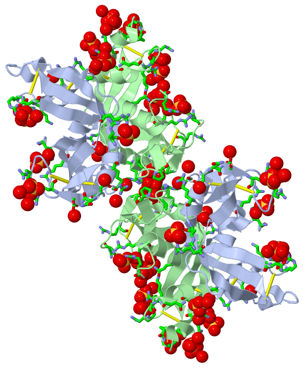
(-) SAPs(SNPs)/Variants  (3, 6)

Asymmetric Unit (3, 6)
  dbSNPPDB
No.SourceVariant IDVariantUniProt IDStatusIDChainVariant
1UniProtVAR_014109R72CECP_HUMANPolymorphism151169198A/BR45C
2UniProtVAR_013149T124RECP_HUMANPolymorphism2073342A/BR97R
3UniProtVAR_029017G130RECP_HUMANPolymorphism12147890A/BG103R

  SNP/SAP Summary Statistics (UniProtKB/Swiss-Prot)
Biological Unit 1 (3, 12)
  dbSNPPDB
No.SourceVariant IDVariantUniProt IDStatusIDChainVariant
1UniProtVAR_014109R72CECP_HUMANPolymorphism151169198A/BR45C
2UniProtVAR_013149T124RECP_HUMANPolymorphism2073342A/BR97R
3UniProtVAR_029017G130RECP_HUMANPolymorphism12147890A/BG103R

  SNP/SAP Summary Statistics (UniProtKB/Swiss-Prot)

(-) PROSITE Motifs  (1, 2)

Asymmetric Unit (1, 2)
 PROSITEUniProtKBPDB
No.IDACDescriptionIDLocationCountLocation
1RNASE_PANCREATICPS00127 Pancreatic ribonuclease family signature.ECP_HUMAN64-70
 
  2A:37-43
B:37-43
Biological Unit 1 (1, 4)
 PROSITEUniProtKBPDB
No.IDACDescriptionIDLocationCountLocation
1RNASE_PANCREATICPS00127 Pancreatic ribonuclease family signature.ECP_HUMAN64-70
 
  4A:37-43
B:37-43

(-) Exons   (1, 2)

Asymmetric Unit (1, 2)
 ENSEMBLUniProtKBPDB
No.Transcript IDExonExon IDGenome LocationLengthIDLocationLengthCountLocationLength
1.1ENST000003046391ENSE00001137146chr14:21359558-2135961053ECP_HUMAN-00--
1.2ENST000003046392ENSE00001124180chr14:21359841-21360507667ECP_HUMAN1-1621622A:1-133
B:1-133
133
133

(-) Sequences/Alignments

Asymmetric Unit
   Reformat: Number of residues per line =  ('0' or empty: single-line sequence representation)
  Number of residues per labelling interval =   
  UniProt sequence: complete  aligned part    
   Show mapping: SCOP domains CATH domains Pfam domains Secondary structure (by author)
SAPs(SNPs) PROSITE motifs Exons
(details for a mapped element are shown in a popup box when the mouse pointer rests over it)
Chain A from PDB  Type:PROTEIN  Length:133
 aligned with ECP_HUMAN | P12724 from UniProtKB/Swiss-Prot  Length:160

    Alignment length:133
                                    37        47        57        67        77        87        97       107       117       127       137       147       157   
            ECP_HUMAN    28 RPPQFTRAQWFAIQHISLNPPRCTIAMRAINNYRWRCKNQNTFLRTTFANVVNVCGNQSIRCPHNRTLNNCHRSRFRVPLLHCDLINPGAQNISNCTYADRPGRRFYVVACDNRDPRDSPRYPVVPVHLDTTI 160
               SCOP domains d4a2oa_ A: Eosinophil cationic protein (ECP), ribonuclease 3                                                                          SCOP domains
               CATH domains ------------------------------------------------------------------------------------------------------------------------------------- CATH domains
               Pfam domains ------------------------------------------------------------------------------------------------------------------------------------- Pfam domains
         Sec.struct. author .....hhhhhhhhhhh.....hhhhhhhhhhh.......eeeee..hhhhhhhhh...ee..........eee....eeeeeeee......hhhhh.eeeeeeee.eeeeeee..........eeeeeeeee. Sec.struct. author
                 SAPs(SNPs) --------------------------------------------C---------------------------------------------------R-----R------------------------------ SAPs(SNPs)
                    PROSITE ------------------------------------RNASE_P------------------------------------------------------------------------------------------ PROSITE
               Transcript 1 Exon 1.2  PDB: A:1-133 UniProt: 1-162 [INCOMPLETE]                                                                                    Transcript 1
                 4a2o A   1 RPPQFTRAQWFAIQHISLNPPRCTIAMRAINNYRWRCKNQNTFLRTTFANVVNVCGNQSIRCPHNRTLNNCHRSRFRVPLLHCDLINPGAQNISNCRYADRPGRRFYVVACDNRDPRDSPRYPVVPVHLDTTI 133
                                    10        20        30        40        50        60        70        80        90       100       110       120       130   

Chain B from PDB  Type:PROTEIN  Length:133
 aligned with ECP_HUMAN | P12724 from UniProtKB/Swiss-Prot  Length:160

    Alignment length:133
                                    37        47        57        67        77        87        97       107       117       127       137       147       157   
            ECP_HUMAN    28 RPPQFTRAQWFAIQHISLNPPRCTIAMRAINNYRWRCKNQNTFLRTTFANVVNVCGNQSIRCPHNRTLNNCHRSRFRVPLLHCDLINPGAQNISNCTYADRPGRRFYVVACDNRDPRDSPRYPVVPVHLDTTI 160
               SCOP domains d4a2ob_ B: Eosinophil cationic protein (ECP), ribonuclease 3                                                                          SCOP domains
               CATH domains ------------------------------------------------------------------------------------------------------------------------------------- CATH domains
               Pfam domains ------------------------------------------------------------------------------------------------------------------------------------- Pfam domains
         Sec.struct. author .....hhhhhhhhhhh.....hhhhhhhhhhh.......eeeee..hhhhhhhhhh..ee..........eee....eeeeeeee......hhhhh.eeeeeeee.eeeeeee..........eeeeeeeee. Sec.struct. author
                 SAPs(SNPs) --------------------------------------------C---------------------------------------------------R-----R------------------------------ SAPs(SNPs)
                    PROSITE ------------------------------------RNASE_P------------------------------------------------------------------------------------------ PROSITE
               Transcript 1 Exon 1.2  PDB: B:1-133 UniProt: 1-162 [INCOMPLETE]                                                                                    Transcript 1
                 4a2o B   1 RPPQFTRAQWFAIQHISLNPPRCTIAMRAINNYRWRCKNQNTFLRTTFANVVNVCGNQSIRCPHNRTLNNCHRSRFRVPLLHCDLINPGAQNISNCRYADRPGRRFYVVACDNRDPRDSPRYPVVPVHLDTTI 133
                                    10        20        30        40        50        60        70        80        90       100       110       120       130   

   Legend:   → Mismatch (orange background)
  - → Gap (green background, '-', border residues have a numbering label)
    → Modified Residue (blue background, lower-case, 'x' indicates undefined single-letter code, labelled with number + name)
  x → Chemical Group (purple background, 'x', labelled with number + name, e.g. ACE or NH2)
  extra numbering lines below/above indicate numbering irregularities and modified residue names etc., number ends below/above '|'

 Classification and Annotation

(-) SCOP Domains  (1, 2)

Asymmetric Unit

(-) CATH Domains  (0, 0)

(no "CATH Domain" information available for 4A2O)

(-) Pfam Domains  (0, 0)

(no "Pfam Domain" information available for 4A2O)

(-) Gene Ontology  (15, 15)

Asymmetric Unit(hide GO term definitions)
Chain A,B   (ECP_HUMAN | P12724)
molecular function
    GO:0004519    endonuclease activity    Catalysis of the hydrolysis of ester linkages within nucleic acids by creating internal breaks.
    GO:0016787    hydrolase activity    Catalysis of the hydrolysis of various bonds, e.g. C-O, C-N, C-C, phosphoric anhydride bonds, etc. Hydrolase is the systematic name for any enzyme of EC class 3.
    GO:0004518    nuclease activity    Catalysis of the hydrolysis of ester linkages within nucleic acids.
    GO:0003676    nucleic acid binding    Interacting selectively and non-covalently with any nucleic acid.
    GO:0004540    ribonuclease activity    Catalysis of the hydrolysis of phosphodiester bonds in chains of RNA.
biological process
    GO:0006401    RNA catabolic process    The chemical reactions and pathways resulting in the breakdown of RNA, ribonucleic acid, one of the two main type of nucleic acid, consisting of a long, unbranched macromolecule formed from ribonucleotides joined in 3',5'-phosphodiester linkage.
    GO:0090501    RNA phosphodiester bond hydrolysis    The RNA metabolic process in which the phosphodiester bonds between ribonucleotides are cleaved by hydrolysis.
    GO:0019731    antibacterial humoral response    An immune response against bacteria mediated through a body fluid. Examples of this process are the antibacterial humoral responses in Mus musculus and Drosophila melanogaster.
    GO:0050830    defense response to Gram-positive bacterium    Reactions triggered in response to the presence of a Gram-positive bacterium that act to protect the cell or organism.
    GO:0042742    defense response to bacterium    Reactions triggered in response to the presence of a bacterium that act to protect the cell or organism.
    GO:0002227    innate immune response in mucosa    Any process of the innate immune response that takes place in the mucosal tissues.
    GO:0090305    nucleic acid phosphodiester bond hydrolysis    The nucleic acid metabolic process in which the phosphodiester bonds between nucleotides are cleaved by hydrolysis.
cellular component
    GO:0070062    extracellular exosome    A vesicle that is released into the extracellular region by fusion of the limiting endosomal membrane of a multivesicular body with the plasma membrane. Extracellular exosomes, also simply called exosomes, have a diameter of about 40-100 nm.
    GO:0005576    extracellular region    The space external to the outermost structure of a cell. For cells without external protective or external encapsulating structures this refers to space outside of the plasma membrane. This term covers the host cell environment outside an intracellular parasite.
    GO:0005615    extracellular space    That part of a multicellular organism outside the cells proper, usually taken to be outside the plasma membranes, and occupied by fluid.

 Visualization

(-) Interactive Views

Asymmetric Unit
  Complete Structure
    Jena3D(integrated viewing of ligand, site, SAP, PROSITE, SCOP information)
    WebMol | AstexViewer[tm]@PDBe
(Java Applets, require no local installation except for Java; loading may be slow)
    STRAP
(Java WebStart application, automatic local installation, requires Java; full application with system access!)
    RasMol
(require local installation)
    Molscript (VRML)
(requires installation of a VRML viewer; select preferred view via VRML and generate a mono or stereo PDF format file)
 
  Ligands, Modified Residues, Ions
    SO4  [ RasMol | Jena3D ]  +environment [ RasMol | Jena3D ]
 
  Sites
    AC1  [ RasMol ]  +environment [ RasMol ]
    AC2  [ RasMol ]  +environment [ RasMol ]
    AC3  [ RasMol ]  +environment [ RasMol ]
    AC4  [ RasMol ]  +environment [ RasMol ]
    AC5  [ RasMol ]  +environment [ RasMol ]
    AC6  [ RasMol ]  +environment [ RasMol ]
    AC7  [ RasMol ]  +environment [ RasMol ]
    AC8  [ RasMol ]  +environment [ RasMol ]
    AC9  [ RasMol ]  +environment [ RasMol ]
    BC1  [ RasMol ]  +environment [ RasMol ]
    BC2  [ RasMol ]  +environment [ RasMol ]
    BC3  [ RasMol ]  +environment [ RasMol ]
    BC4  [ RasMol ]  +environment [ RasMol ]
    BC5  [ RasMol ]  +environment [ RasMol ]
 
  Cis Peptide Bonds
(no "Cis Peptide Bonds" information available for 4a2o)
 
Biological Unit
  Complete Structure
    Biological Unit 1  [ Jena3D ]

(-) Still Images

Jmol
  protein: cartoon or spacefill or dots and stick; nucleic acid: cartoon and stick; ligands: spacefill; active site: stick
Molscript
  protein, nucleic acid: cartoon; ligands: spacefill; active site: ball and stick

 Databases and Analysis Tools

(-) Databases

Access by PDB/NDB ID
  4a2o
    Family and Domain InformationProDom | SYSTERS
    General Structural InformationGlycoscienceDB | MMDB | NDB | OCA | PDB | PDBe | PDBj | PDBsum | PDBWiki | PQS | PROTEOPEDIA
    Orientation in MembranesOPM
    Protein SurfaceSURFACE
    Secondary StructureDSSP (structure derived) | HSSP (homology derived)
    Structural GenomicsGeneCensus
    Structural NeighboursCE | VAST
    Structure ClassificationCATH | Dali | SCOP
    Validation and Original DataBMRB Data View | BMRB Restraints Grid | EDS | PROCHECK | RECOORD | WHAT_CHECK
 
Access by UniProt ID/Accession number
  ECP_HUMAN | P12724
    Comparative Protein Structure ModelsModBase
    Genomic InformationEnsembl
    Protein-protein InteractionDIP
    Sequence, Family and Domain InformationInterPro | Pfam | SMART | UniProtKB/SwissProt
 
Access by Enzyme Classificator   (EC Number)
  3.1.27.-
    General Enzyme InformationBRENDA | EC-PDB | Enzyme | IntEnz
    PathwayKEGG | MetaCyc
 
Access by Disease Identifier   (MIM ID)
  (no 'MIM ID' available)
    Disease InformationOMIM
 
Access by GenAge ID
  (no 'GenAge ID' available)
    Age Related InformationGenAge

(-) Analysis Tools

Access by PDB/NDB ID
    Domain InformationXDom
    Interatomic Contacts of Structural UnitsCSU
    Ligand-protein ContactsLPC
    Protein CavitiescastP
    Sequence and Secondary StructurePDBCartoon
    Structure AlignmentSTRAP(Java WebStart application, automatic local installation, requires Java; full application with system access!)
    Structure and Sequence BrowserSTING
 
Access by UniProt ID/Accession number
  ECP_HUMAN | P12724
    Protein Disorder PredictionDisEMBL | FoldIndex | GLOBPLOT (for more information see DisProt)

 Related Entries

(-) Entries Sharing at Least One Protein Chain (UniProt ID)

UniProtKB/Swiss-Prot
        ECP_HUMAN | P127241dyt 1h1h 1qmt 2kb5 2lvz 4a2y 4owz 4oxb 4oxf 4x08

(-) Related Entries Specified in the PDB File

1dyt X-RAY CRYSTAL STRUCTURE OF ECP (RNASE 3) AT 1.75 A
1h1h CRYSTAL STRUCTURE OF EOSINOPHIL CATIONIC PROTEIN IN COMPLEX WITH 2',5'-ADP AT 2.0 A RESOLUTION REVEALS THE DETAILS OF THE RIBONUCLEOLYTIC ACTIVE SITE
1qmt RECOMBINANT HUMAN EOSINOPHIL CATIONIC PROTEIN
4a2y STRUCTURE OF THE HUMAN EOSINOPHIL CATIONIC PROTEIN IN COMPLEX WITH CITRATE ANIONS