Show PDB file:   
         Plain Text   HTML   (compressed file size)
QuickSearch:   
by PDB,NDB,UniProt,PROSITE Code or Search Term(s)  
(-)Asym./Biol. Unit
(-)Asym./Biol. Unit - sites
collapse expand < >
Image Asym./Biol. Unit
Asym./Biol. Unit  (Jmol Viewer)
Image Asym./Biol. Unit - sites
Asym./Biol. Unit - sites  (Jmol Viewer)

(-) Description

Title :  HUMAN GAR TRANSFORMYLASE IN COMPLEX WITH GAR AND PEMETREXED
 
Authors :  S. M. Deis, C. E. Dann Iii
Date :  22 May 15  (Deposition) - 16 Dec 15  (Release) - 16 Dec 15  (Revision)
Method :  X-RAY DIFFRACTION
Resolution :  2.50
Chains :  Asym./Biol. Unit :  A
Keywords :  Gar Transformylase, Antifolate, Transferase-Transferase Inhibitor Complex (Keyword Search: [Gene Ontology, PubMed, Web (Google))
 
Reference :  L. Wang, A. Wallace, S. Raghavan, S. M. Deis, M. R. Wilson, S. Yang, L. Polin, K. White, J. Kushner, S. Orr, C. George, C. O'Connor, Z. Hou, S. Mitchell-Ryan, C. E. Dann, L. H. Matherly, A. Gangjee
6-Substituted Pyrrolo[2, 3-D]Pyrimidine Thienoyl Regioisomer As Targeted Antifolates For Folate Receptor Alpha And The Proton-Coupled Folate Transporter In Human Tumors.
J. Med. Chem. V. 58 6938 2015
PubMed-ID: 26317331  |  Reference-DOI: 10.1021/ACS.JMEDCHEM.5B00801

(-) Compounds

Molecule 1 - TRIFUNCTIONAL PURINE BIOSYNTHETIC PROTEIN ADENOSINE-3
    ChainsA
    EC Number6.3.4.13, 6.3.3.1, 2.1.2.2
    EngineeredYES
    Expression SystemESCHERICHIA COLI
    Expression System PlasmidPET22B
    Expression System StrainROSETTA(DE3)PLYSS
    Expression System Taxid562
    Expression System Vector TypePLASMID
    FragmentUNP RESIDUES 808-1010
    GeneGART, PGFT, PRGS
    Organism CommonHUMAN
    Organism ScientificHOMO SAPIENS
    Organism Taxid9606

 Structural Features

(-) Chains, Units

  1
Asymmetric/Biological Unit A

Summary Information (see also Sequences/Alignments below)
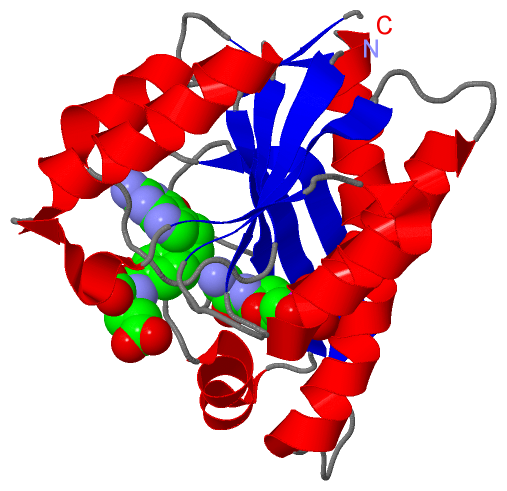
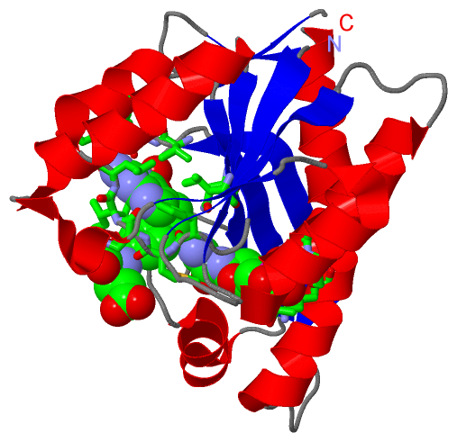
(-) Ligands, Modified Residues, Ions  (2, 2)

Asymmetric/Biological Unit (2, 2)
No.NameCountTypeFull Name
14DW1Ligand/IonN-{4-[2-(2-AMINO-4-OXO-4,7-DIHYDRO-3H-PYRROLO[2,3-D]PYRIMIDIN-5-YL)ETHYL]BENZOYL}-L-GLUTAMIC ACID
2GAR1Ligand/IonGLYCINAMIDE RIBONUCLEOTIDE

(-) Sites  (2, 2)

Asymmetric Unit (2, 2)
No.NameEvidenceResiduesDescription
1AC1SOFTWARELEU A:892 , PHE A:895 , MET A:896 , ARG A:897 , ILE A:898 , LEU A:899 , VAL A:904 , ASN A:913 , VAL A:946 , ALA A:947 , GLU A:948 , VAL A:950 , ASP A:951 , GAR A:1102 , HOH A:1216binding site for residue 4DW A 1101
2AC2SOFTWARETHR A:817 , GLY A:818 , SER A:819 , ASN A:820 , PRO A:916 , LYS A:977 , GLU A:980 , 4DW A:1101binding site for residue GAR A 1102

(-) SS Bonds  (0, 0)

(no "SS Bond" information available for 4ZZ3)

(-) Cis Peptide Bonds  (1, 1)

Asymmetric/Biological Unit
No.Residues
1Leu A:919 -Pro A:920

 Sequence-Structure Mapping

(-) SAPs(SNPs)/Variants  (0, 0)

(no "SAP(SNP)/Variant" information available for 4ZZ3)

(-) PROSITE Motifs  (0, 0)

(no "PROSITE Motif" information available for 4ZZ3)

(-) Exons   (0, 0)

(no "Exon" information available for 4ZZ3)

(-) Sequences/Alignments

Asymmetric/Biological Unit
   Reformat: Number of residues per line =  ('0' or empty: single-line sequence representation)
  Number of residues per labelling interval =   
  UniProt sequence: complete  aligned part    
   Show mapping: SCOP domains CATH domains Pfam domains Secondary structure (by author)
SAPs(SNPs) PROSITE motifs Exons
(details for a mapped element are shown in a popup box when the mouse pointer rests over it)
Chain A from PDB  Type:PROTEIN  Length:200
                                                                                                                                                                                                                                         
               SCOP domains -------------------------------------------------------------------------------------------------------------------------------------------------------------------------------------------------------- SCOP domains
               CATH domains -------------------------------------------------------------------------------------------------------------------------------------------------------------------------------------------------------- CATH domains
               Pfam domains -------------------------------------------------------------------------------------------------------------------------------------------------------------------------------------------------------- Pfam domains
         Sec.struct. author .eeeeee...hhhhhhhhhhhh......eeeeeee....hhhhhhhhhhh..eee.hhhhh.hhhhhhhhhhhhhhhh...eeee.......hhhhhhhh...eeeee..........hhhhhhhhhh..eeeeeeee.........eeeeeeee.....hhhhhhhhhhhhhhhhhhhhhhhhhh..eee.....eee. Sec.struct. author
                 SAPs(SNPs) -------------------------------------------------------------------------------------------------------------------------------------------------------------------------------------------------------- SAPs(SNPs)
                    PROSITE -------------------------------------------------------------------------------------------------------------------------------------------------------------------------------------------------------- PROSITE
                 Transcript -------------------------------------------------------------------------------------------------------------------------------------------------------------------------------------------------------- Transcript
                4zz3 A  808 ARVAVLISGTGSNLQALIDSTREPNSSAQIDIVISNKAAVAGLDKAERAGIPTRVINHKLYKNRVEFDSAIDLVLEEFSIDIVCLAGFMRILSGPFVQKWNGKMLNIHPSLLPSFKGSNAHEQALETGVTVTGCTVHFVAEDVDAGQIILQEAVPVKRGDTVATLSERVKLAEHKIFPAALQLVASGTVQLGENGKICWV 1007
                                   817       827       837       847       857       867       877       887       897       907       917       927       937       947       957       967       977       987       997      1007

   Legend:   → Mismatch (orange background)
  - → Gap (green background, '-', border residues have a numbering label)
    → Modified Residue (blue background, lower-case, 'x' indicates undefined single-letter code, labelled with number + name)
  x → Chemical Group (purple background, 'x', labelled with number + name, e.g. ACE or NH2)
  extra numbering lines below/above indicate numbering irregularities and modified residue names etc., number ends below/above '|'

 Classification and Annotation

(-) SCOP Domains  (0, 0)

(no "SCOP Domain" information available for 4ZZ3)

(-) CATH Domains  (0, 0)

(no "CATH Domain" information available for 4ZZ3)

(-) Pfam Domains  (0, 0)

(no "Pfam Domain" information available for 4ZZ3)

(-) Gene Ontology  (27, 27)

Asymmetric/Biological Unit(hide GO term definitions)

 Visualization

(-) Interactive Views

Asymmetric/Biological Unit
  Complete Structure
    Jena3D(integrated viewing of ligand, site, SAP, PROSITE, SCOP information)
    WebMol | AstexViewer[tm]@PDBe
(Java Applets, require no local installation except for Java; loading may be slow)
    STRAP
(Java WebStart application, automatic local installation, requires Java; full application with system access!)
    RasMol
(require local installation)
    Molscript (VRML)
(requires installation of a VRML viewer; select preferred view via VRML and generate a mono or stereo PDF format file)
 
  Ligands, Modified Residues, Ions
    4DW  [ RasMol | Jena3D ]  +environment [ RasMol | Jena3D ]
    GAR  [ RasMol | Jena3D ]  +environment [ RasMol | Jena3D ]
 
  Sites
    AC1  [ RasMol ]  +environment [ RasMol ]
    AC2  [ RasMol ]  +environment [ RasMol ]
 
  Cis Peptide Bonds
    Leu A:919 - Pro A:920   [ RasMol ]  
 

(-) Still Images

Jmol
  protein: cartoon or spacefill or dots and stick; nucleic acid: cartoon and stick; ligands: spacefill; active site: stick
Molscript
  protein, nucleic acid: cartoon; ligands: spacefill; active site: ball and stick

 Databases and Analysis Tools

(-) Databases

Access by PDB/NDB ID
  4zz3
    Family and Domain InformationProDom | SYSTERS
    General Structural InformationGlycoscienceDB | MMDB | NDB | OCA | PDB | PDBe | PDBj | PDBsum | PDBWiki | PQS | PROTEOPEDIA
    Orientation in MembranesOPM
    Protein SurfaceSURFACE
    Secondary StructureDSSP (structure derived) | HSSP (homology derived)
    Structural GenomicsGeneCensus
    Structural NeighboursCE | VAST
    Structure ClassificationCATH | Dali | SCOP
    Validation and Original DataBMRB Data View | BMRB Restraints Grid | EDS | PROCHECK | RECOORD | WHAT_CHECK
 
Access by UniProt ID/Accession number
  PUR2_HUMAN | P22102
    Comparative Protein Structure ModelsModBase
    Genomic InformationEnsembl
    Protein-protein InteractionDIP
    Sequence, Family and Domain InformationInterPro | Pfam | SMART | UniProtKB/SwissProt
 
Access by Enzyme Classificator   (EC Number)
  2.1.2.2
    General Enzyme InformationBRENDA | EC-PDB | Enzyme | IntEnz
    PathwayKEGG | MetaCyc
  6.3.3.1
    General Enzyme InformationBRENDA | EC-PDB | Enzyme | IntEnz
    PathwayKEGG | MetaCyc
  6.3.4.13
    General Enzyme InformationBRENDA | EC-PDB | Enzyme | IntEnz
    PathwayKEGG | MetaCyc
 
Access by Disease Identifier   (MIM ID)
  (no 'MIM ID' available)
    Disease InformationOMIM
 
Access by GenAge ID
  (no 'GenAge ID' available)
    Age Related InformationGenAge

(-) Analysis Tools

Access by PDB/NDB ID
    Domain InformationXDom
    Interatomic Contacts of Structural UnitsCSU
    Ligand-protein ContactsLPC
    Protein CavitiescastP
    Sequence and Secondary StructurePDBCartoon
    Structure AlignmentSTRAP(Java WebStart application, automatic local installation, requires Java; full application with system access!)
    Structure and Sequence BrowserSTING
 
Access by UniProt ID/Accession number
  PUR2_HUMAN | P22102
    Protein Disorder PredictionDisEMBL | FoldIndex | GLOBPLOT (for more information see DisProt)

 Related Entries

(-) Entries Sharing at Least One Protein Chain (UniProt ID)

UniProtKB/Swiss-Prot
        PUR2_HUMAN | P221021mej 1men 1meo 1njs 1rbm 1rbq 1rby 1rbz 1rc0 1rc1 1zlx 1zly 2qk4 2v9y 4ew1 4ew2 4ew3 4zyt 4zyu 4zyv 4zyw 4zyx 4zyy 4zyz 4zz0 4zz1 4zz2 5j9f

(-) Related Entries Specified in the PDB File

1mej 1MEJ IS THE SAME PROTEIN IN APO FORM AT PH 8.5
1men 1MEN IS THE SAME PROTEIN COMPLEXED WITH BETA-GAR
1meo 1MEO IS THE SAME PROTEIN IN APO FORM AT PH 4.2
1njs 1NJS IS THE SAME PROTEIN COMPLEXED WITH A HYDROLYZED FORM OF 10- (TRIFLUOROACETYL)-5,10-DIDEAZAACYCLIC-5,6,7,8-TETRAHYDROFOLIC ACID
1rbm 1RBM IS THE SAME PROTEIN COMPLEXED WITH POLYGLUTAMINATED 10- (TRIFLUOROACETYL)-5,10-DIDEAZAACYCLIC-5,6,7,8-TETRAHYDROFOLIC ACID
1rbq 1RBQ IS THE SAME PROTEIN COMPLEXED WITH 10-(TRIFLUOROACETYL)-5,10- DIDEAZAACYCLIC-5,6,7,8-TETRAHYDROFOLIC ACID
1rby 1RBY IS THE SAME PROTEIN COMPLEXED WITH 10-(TRIFLUOROACETYL)-5,10- DIDEAZAACYCLIC-5,6,7,8-TETRAHYDROFOLIC ACID AND BETA-GAR
1rbz 1RBZ IS THE SAME PROTEIN COMPLEXED WITH POLYGLUTAMINATED 10- (TRIFLUOROACETYL)-5,10-DIDEAZAACYCLIC-5,6,7,8-TETRAHYDROFOLIC ACID
1rc0 1RC0 IS THE SAME PROTEIN COMPLEXED WITH POLYGLUTAMINATED 10- (TRIFLUOROACETYL)-5,10-DIDEAZAACYCLIC-5,6,7,8-TETRAHYDROFOLIC ACID
1rc1 1RC1 IS THE SAME PROTEIN COMPLEXED WITH POLYGLUTAMINATED 10- (TRIFLUOROACETYL)-5,10-DIDEAZAACYCLIC-5,6,7,8-TETRAHYDROFOLIC ACID
1zlx 1ZLX IS THE SAME PROTEIN IN APO FORM
1zly 1ZLY IS THE SAME PROTEIN COMPLEXED WITH 4-[(4-{[(2-AMINO-4-OXO-3,4- DIHYDROQUINAZOLIN- 6-YL)METHYL]AMINO}BENZOYL)AMINO]BUTANOIC ACID AND 5-O-PHOSPHONO-BETA-D-RIBOFURANOSYLAMINE
4ew2 4EW2 IS THE SAME PROTEIN COMPLEXED WITH 10S-METHYLTHIO-DDATHF
4ew3 4EW3 IS THE SAME PROTEIN COMPLEXED WITH 10R-METHYLTHIO-DDATHF
4zyt
4zyu
4zyv
4zyw
4zyx
4zyy
4zyz
4zz0
4zz1
4zz2