Show PDB file:   
         Plain Text   HTML   (compressed file size)
QuickSearch:   
by PDB,NDB,UniProt,PROSITE Code or Search Term(s)  
(-)Asymmetric Unit
(-)Asym. Unit - sites
(-)Biological Unit 1
(-)Biol. Unit 1 - sites
(-)Biological Unit 2
collapse expand < >
Image Asymmetric Unit
Asymmetric Unit  (Jmol Viewer)
Image Asym. Unit - sites
Asym. Unit - sites  (Jmol Viewer)
Image Biological Unit 1
Biological Unit 1  (Jmol Viewer)
Image Biol. Unit 1 - sites
Biol. Unit 1 - sites  (Jmol Viewer)
Image Biological Unit 2
Biological Unit 2  (Jmol Viewer)

(-) Description

Title :  STRUCTURE OF CFA/I PILI MAJOR SUBUNIT CFAB TRIMER
 
Authors :  R. Bao, D. Xia
Date :  10 Feb 15  (Deposition) - 10 Aug 16  (Release) - 10 Aug 16  (Revision)
Method :  X-RAY DIFFRACTION
Resolution :  2.40
Chains :  Asym. Unit :  A,B,C
Biol. Unit 1:  A,B  (3x)
Biol. Unit 2:  C  (6x)
Keywords :  Enterotoxigenic Escherichia Coli, Periplasmic Chaperone, Major Pilin, Self-Assembly, Fimbriae, Structural Protein (Keyword Search: [Gene Ontology, PubMed, Web (Google))
 
Reference :  R. Bao, S. J. Savarino, D. Xia
Periplasmic Chaperone Cfaa Of Cfa/I Fimbriae Functions To Maintain Assembly Potency And Prevent Self-Polymerization O Pilin Subunits
To Be Published
PubMed: search

(-) Compounds

Molecule 1 - CFA/I FIMBRIAL SUBUNIT B
    ChainsB, A, C
    EngineeredYES
    Expression SystemESCHERICHIA COLI
    Expression System Taxid562
    FragmentUNP RESIDUES 25-170
    GeneCFAB, ETEC_P948_0400
    Organism Taxid316401
    StrainH10407 / ETEC
    SynonymCFA/I ANTIGEN,CFA/I PILIN,COLONIZATION FACTOR ANTIGEN I SUBUNIT B

 Structural Features

(-) Chains, Units

  123
Asymmetric Unit ABC
Biological Unit 1 (3x)AB 
Biological Unit 2 (6x)  C

Summary Information (see also Sequences/Alignments below)

(-) Ligands, Modified Residues, Ions  (2, 4)

Asymmetric Unit (2, 4)
No.NameCountTypeFull Name
1EDO1Ligand/Ion1,2-ETHANEDIOL
2MG3Ligand/IonMAGNESIUM ION
Biological Unit 1 (1, 3)
No.NameCountTypeFull Name
1EDO3Ligand/Ion1,2-ETHANEDIOL
2MG-1Ligand/IonMAGNESIUM ION
Biological Unit 2 (0, 0)
No.NameCountTypeFull Name
1EDO-1Ligand/Ion1,2-ETHANEDIOL
2MG-1Ligand/IonMAGNESIUM ION

(-) Sites  (2, 2)

Asymmetric Unit (2, 2)
No.NameEvidenceResiduesDescription
1AC1SOFTWAREASP B:21 , ASN B:23 , GLU B:41 , SER B:42 , HOH B:368binding site for residue EDO B 202
2AC2SOFTWAREILE B:80 , LEU B:89binding site for residue MG B 203

(-) SS Bonds  (0, 0)

(no "SS Bond" information available for 4Y2N)

(-) Cis Peptide Bonds  (0, 0)

(no "Cis Peptide Bond" information available for 4Y2N)

 Sequence-Structure Mapping

(-) SAPs(SNPs)/Variants  (0, 0)

(no "SAP(SNP)/Variant" information available for 4Y2N)

(-) PROSITE Motifs  (0, 0)

(no "PROSITE Motif" information available for 4Y2N)

(-) Exons   (0, 0)

(no "Exon" information available for 4Y2N)

(-) Sequences/Alignments

Asymmetric Unit
   Reformat: Number of residues per line =  ('0' or empty: single-line sequence representation)
  Number of residues per labelling interval =   
  UniProt sequence: complete  aligned part    
   Show mapping: SCOP domains CATH domains Pfam domains Secondary structure (by author)
SAPs(SNPs) PROSITE motifs Exons
(details for a mapped element are shown in a popup box when the mouse pointer rests over it)
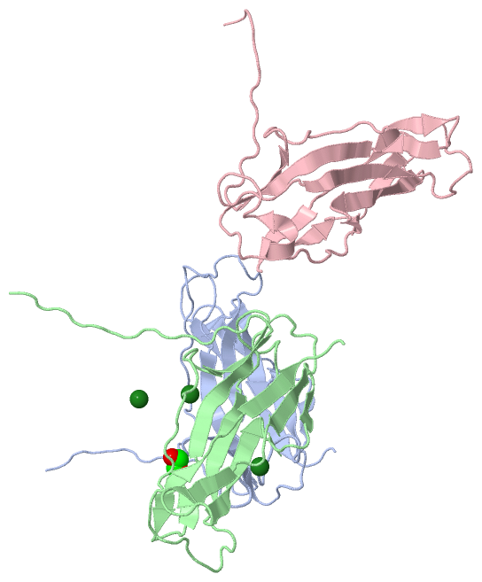
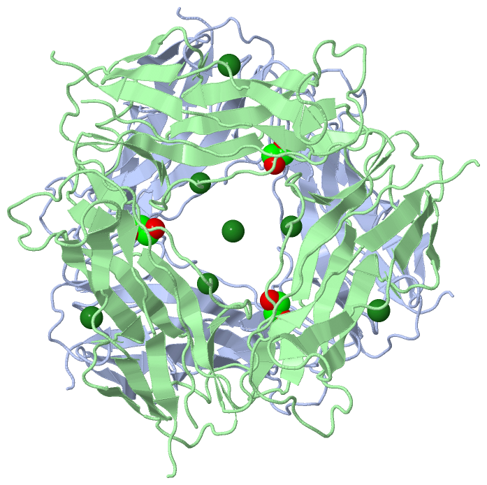
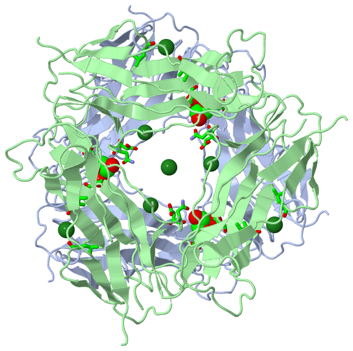
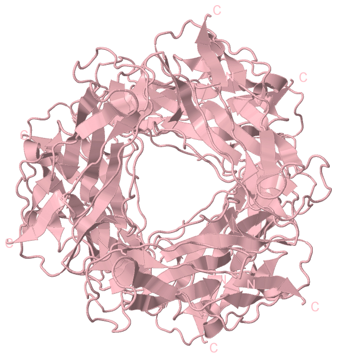
Chain A from PDB  Type:PROTEIN  Length:147
                                                                                                                                                                                   
               SCOP domains --------------------------------------------------------------------------------------------------------------------------------------------------- SCOP domains
               CATH domains --------------------------------------------------------------------------------------------------------------------------------------------------- CATH domains
               Pfam domains --------------------------------------------------------------------------------------------------------------------------------------------------- Pfam domains
         Sec.struct. author ..............eeeee.......................eeeeeeeee......eeee.....eeee......eeeeeeee..eee....eee..................eeeeeee.............ee.....eee... Sec.struct. author
                 SAPs(SNPs) --------------------------------------------------------------------------------------------------------------------------------------------------- SAPs(SNPs)
                    PROSITE --------------------------------------------------------------------------------------------------------------------------------------------------- PROSITE
                 Transcript --------------------------------------------------------------------------------------------------------------------------------------------------- Transcript
                 4y2n A   1 HEKNITVTASVDPVIDLLQADGNALPSAVKLAYSPASKTFESYRVMTQVHTNDATKKVIVKLADTPQLTDVLNSTVQMPISVSWGGQVLSTTAKEFEAAALGYSASGVNGVSSSQELVISAAPKTAGTAPTAGNYSGVVSLVMTLGS 147
                                    10        20        30        40        50        60        70        80        90       100       110       120       130       140       

Chain B from PDB  Type:PROTEIN  Length:149
                                                                                                                                                                                     
               SCOP domains ----------------------------------------------------------------------------------------------------------------------------------------------------- SCOP domains
               CATH domains ----------------------------------------------------------------------------------------------------------------------------------------------------- CATH domains
               Pfam domains ----------------------------------------------------------------------------------------------------------------------------------------------------- Pfam domains
         Sec.struct. author ................eeeee.............ee....ee..eeeeeeeee......eeeee....eeee......eeeeeeee..ee.....eee..................eeeeeee.............ee....eeee... Sec.struct. author
                 SAPs(SNPs) ----------------------------------------------------------------------------------------------------------------------------------------------------- SAPs(SNPs)
                    PROSITE ----------------------------------------------------------------------------------------------------------------------------------------------------- PROSITE
                 Transcript ----------------------------------------------------------------------------------------------------------------------------------------------------- Transcript
                 4y2n B  -1 HHHEKNITVTASVDPVIDLLQADGNALPSAVKLAYSPASKTFESYRVMTQVHTNDATKKVIVKLADTPQLTDVLNSTVQMPISVSWGGQVLSTTAKEFEAAALGYSASGVNGVSSSQELVISAAPKTAGTAPTAGNYSGVVSLVMTLGS 147
                                     8        18        28        38        48        58        68        78        88        98       108       118       128       138         

Chain C from PDB  Type:PROTEIN  Length:147
                                                                                                                                                                                   
               SCOP domains --------------------------------------------------------------------------------------------------------------------------------------------------- SCOP domains
               CATH domains --------------------------------------------------------------------------------------------------------------------------------------------------- CATH domains
               Pfam domains --------------------------------------------------------------------------------------------------------------------------------------------------- Pfam domains
         Sec.struct. author ...............eeeee............eee....eee.eeeeeeeee......eeee.....eeee......eeeeeeee..eee........hhhhh............eeeeeee.............ee.....eeee. Sec.struct. author
                 SAPs(SNPs) --------------------------------------------------------------------------------------------------------------------------------------------------- SAPs(SNPs)
                    PROSITE --------------------------------------------------------------------------------------------------------------------------------------------------- PROSITE
                 Transcript --------------------------------------------------------------------------------------------------------------------------------------------------- Transcript
                 4y2n C   0 HHEKNITVTASVDPVIDLLQADGNALPSAVKLAYSPASKTFESYRVMTQVHTNDATKKVIVKLADTPQLTDVLNSTVQMPISVSWGGQVLSTTAKEFEAAALGYSASGVNGVSSSQELVISAAPKTAGTAPTAGNYSGVVSLVMTLG 146
                                     9        19        29        39        49        59        69        79        89        99       109       119       129       139       

   Legend:   → Mismatch (orange background)
  - → Gap (green background, '-', border residues have a numbering label)
    → Modified Residue (blue background, lower-case, 'x' indicates undefined single-letter code, labelled with number + name)
  x → Chemical Group (purple background, 'x', labelled with number + name, e.g. ACE or NH2)
  extra numbering lines below/above indicate numbering irregularities and modified residue names etc., number ends below/above '|'

 Classification and Annotation

(-) SCOP Domains  (0, 0)

(no "SCOP Domain" information available for 4Y2N)

(-) CATH Domains  (0, 0)

(no "CATH Domain" information available for 4Y2N)

(-) Pfam Domains  (0, 0)

(no "Pfam Domain" information available for 4Y2N)

(-) Gene Ontology  (1, 1)

Asymmetric Unit(hide GO term definitions)

 Visualization

(-) Interactive Views

Asymmetric Unit
  Complete Structure
    Jena3D(integrated viewing of ligand, site, SAP, PROSITE, SCOP information)
    WebMol | AstexViewer[tm]@PDBe
(Java Applets, require no local installation except for Java; loading may be slow)
    STRAP
(Java WebStart application, automatic local installation, requires Java; full application with system access!)
    RasMol
(require local installation)
    Molscript (VRML)
(requires installation of a VRML viewer; select preferred view via VRML and generate a mono or stereo PDF format file)
 
  Ligands, Modified Residues, Ions
    EDO  [ RasMol | Jena3D ]  +environment [ RasMol | Jena3D ]
    MG  [ RasMol | Jena3D ]  +environment [ RasMol | Jena3D ]
 
  Sites
    AC1  [ RasMol ]  +environment [ RasMol ]
    AC2  [ RasMol ]  +environment [ RasMol ]
 
  Cis Peptide Bonds
(no "Cis Peptide Bonds" information available for 4y2n)
 
Biological Units
  Complete Structure
    Biological Unit 1  [ Jena3D ]
    Biological Unit 2  [ Jena3D ]

(-) Still Images

Jmol
  protein: cartoon or spacefill or dots and stick; nucleic acid: cartoon and stick; ligands: spacefill; active site: stick
Molscript
  protein, nucleic acid: cartoon; ligands: spacefill; active site: ball and stick

 Databases and Analysis Tools

(-) Databases

Access by PDB/NDB ID
  4y2n
    Family and Domain InformationProDom | SYSTERS
    General Structural InformationGlycoscienceDB | MMDB | NDB | OCA | PDB | PDBe | PDBj | PDBsum | PDBWiki | PQS | PROTEOPEDIA
    Orientation in MembranesOPM
    Protein SurfaceSURFACE
    Secondary StructureDSSP (structure derived) | HSSP (homology derived)
    Structural GenomicsGeneCensus
    Structural NeighboursCE | VAST
    Structure ClassificationCATH | Dali | SCOP
    Validation and Original DataBMRB Data View | BMRB Restraints Grid | EDS | PROCHECK | RECOORD | WHAT_CHECK
 
Access by UniProt ID/Accession number
  FMC1_ECOH1 | E3PPC4
    Comparative Protein Structure ModelsModBase
    Genomic InformationEnsembl
    Protein-protein InteractionDIP
    Sequence, Family and Domain InformationInterPro | Pfam | SMART | UniProtKB/SwissProt
 
Access by Enzyme Classificator   (EC Number)
  (no 'Enzyme Classificator' available)
    General Enzyme InformationBRENDA | EC-PDB | Enzyme | IntEnz
    PathwayKEGG | MetaCyc
 
Access by Disease Identifier   (MIM ID)
  (no 'MIM ID' available)
    Disease InformationOMIM
 
Access by GenAge ID
  (no 'GenAge ID' available)
    Age Related InformationGenAge

(-) Analysis Tools

Access by PDB/NDB ID
    Domain InformationXDom
    Interatomic Contacts of Structural UnitsCSU
    Ligand-protein ContactsLPC
    Protein CavitiescastP
    Sequence and Secondary StructurePDBCartoon
    Structure AlignmentSTRAP(Java WebStart application, automatic local installation, requires Java; full application with system access!)
    Structure and Sequence BrowserSTING
 
Access by UniProt ID/Accession number
  FMC1_ECOH1 | E3PPC4
    Protein Disorder PredictionDisEMBL | FoldIndex | GLOBPLOT (for more information see DisProt)

 Related Entries

(-) Entries Sharing at Least One Protein Chain (UniProt ID)

UniProtKB/Swiss-Prot
        FMC1_ECOH1 | E3PPC43f83 3f84 3f85 4y2l 4y2o

(-) Related Entries Specified in the PDB File

4y2l 4y2o