|
|
|
|
 Description
|
|
Compounds
|
||||||||||||||||||||||||||||||||||||||||||||||||||||||||||||||||||||||
Chains, Units
Summary Information (see also Sequences/Alignments below) |
Ligands, Modified Residues, Ions (8, 47)
Asymmetric/Biological Unit (8, 47)
|
Sites (8, 8)
Asymmetric Unit (8, 8)
|
SS Bonds (2, 2)
Asymmetric/Biological Unit
|
||||||||||||
Cis Peptide Bonds (0, 0)| (no "Cis Peptide Bond" information available for 4R8I) |
SAPs(SNPs)/Variants (0, 0)| (no "SAP(SNP)/Variant" information available for 4R8I) |
PROSITE Motifs (0, 0)| (no "PROSITE Motif" information available for 4R8I) |
Exons (0, 0)| (no "Exon" information available for 4R8I) |
Sequences/Alignments
Asymmetric/Biological Unit
Chain A from PDB Type:PROTEIN Length:68
SCOP domains -------------------------------------------------------------------- SCOP domains
CATH domains -------------------------------------------------------------------- CATH domains
Pfam domains -------------------------------------------------------------------- Pfam domains
SAPs(SNPs) -------------------------------------------------------------------- SAPs(SNPs)
PROSITE -------------------------------------------------------------------- PROSITE
Transcript -------------------------------------------------------------------- Transcript
4r8i A 3 DAINAPVTCCYNFTNRKISVQRLASYRRITSSKCPKEAVIFKTIVAKEICADPKQKWVQDSMDHLDKQ 70
12 22 32 42 52 62
Chain B from PDB  Type:OTHER  Length:40

                  4r8i B  1 xxxxxxxxxxxxxxxxxxxxxxxxxxxxxxxxxxxxxxxx 40
                            ||||||||10||||||||20||||||||30||||||||40
                            1-0G||||10-0U||||19-0C||||28-0C||||37-0U
                             2-0C||||11-0C||||20-0A||||29-0C||||38-0G
                              3-0A||| 12-0A||| 21-0A||| 30-0G||| 39-0C
                               4-0C||  13-0C||  22-0G||  31-0U1|  40-0G
                                5-0G|   14-0C|   23-0U|   32-0G|
                                 6-0U    15-0G    24-0G    33-0G
                                  7-0C    16-0G    25-0A    34-0C
                                   8-0C    17-0U    26-0A    35-0U
                                    9-0C    18-0G    27-0G    36-0C
|
||||||||||||||||||||
SCOP Domains (0, 0)| (no "SCOP Domain" information available for 4R8I) |
CATH Domains (0, 0)| (no "CATH Domain" information available for 4R8I) |
Pfam Domains (0, 0)| (no "Pfam Domain" information available for 4R8I) |
Gene Ontology (113, 113)|
Asymmetric/Biological Unit(hide GO term definitions) |
Interactive Views
|
||||||||||||||||||||||||||||||||||||||||||||||||||||||||||||||||||||||||||||||||||||||||||||||||||||||||||||||||||||||||||||||||||||||||||||||||||||||||||||||||||||||||||||||||||||||||||||||||||||||||||||||||||||||||
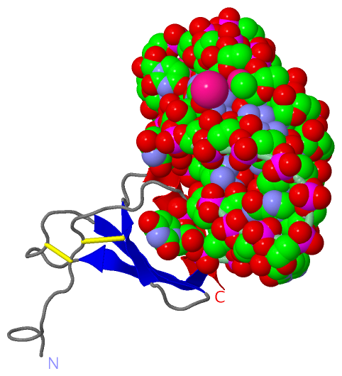
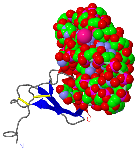
Still Images
|
||||||||||||||||
Databases
|
||||||||||||||||||||||||||||||||||||||||||||||||||||||||||||||||||||||||||||||||||||||||||||||||||||||||||||||||||||||||||||||||||||||||||||||||||||||||||||||||
Analysis Tools
|
|||||||||||||||||||||||||||||||||||||||||||||||||||||||||||||
Entries Sharing at Least One Protein Chain (UniProt ID)
Related Entries Specified in the PDB File
|
|