Show PDB file:   
         Plain Text   HTML   (compressed file size)
QuickSearch:   
by PDB,NDB,UniProt,PROSITE Code or Search Term(s)  
(-)Asymmetric Unit
(-)Asym. Unit - sites
(-)Biological Unit 1
(-)Biol. Unit 1 - sites
(-)Biological Unit 2
collapse expand < >
Image Asymmetric Unit
Asymmetric Unit  (Jmol Viewer)
Image Asym. Unit - sites
Asym. Unit - sites  (Jmol Viewer)
Image Biological Unit 1
Biological Unit 1  (Jmol Viewer)
Image Biol. Unit 1 - sites
Biol. Unit 1 - sites  (Jmol Viewer)
Image Biological Unit 2
Biological Unit 2  (Jmol Viewer)

(-) Description

Title :  THE CRYSTAL STRUCTURE OF R60E MUTANT OF THE HISTIDINE KINASE (KINB) SENSOR DOMAIN FROM PSEUDOMONAS AERUGINOSA PA01
 
Authors :  K. Tan, G. Chhor, R. Jedrzejczak, A. Joachimiak, Midwest Center For Structural Genomics (Mcsg)
Date :  09 Jul 13  (Deposition) - 07 Aug 13  (Release) - 14 Aug 13  (Revision)
Method :  X-RAY DIFFRACTION
Resolution :  2.00
Chains :  Asym. Unit :  A,B
Biol. Unit 1:  A  (1x)
Biol. Unit 2:  B  (1x)
Keywords :  Structural Genomics, Psi-Biology, Protein Structure Initiative, Midwest Center For Structural Genomics, Mcsg, Transferase (Keyword Search: [Gene Ontology, PubMed, Web (Google))
 
Reference :  K. Tan, G. Chhor, R. Jedrzejczak, A. Joachimiak
The Crystal Structure Of R60E Mutant Of The Histidine Kinas (Kinb) Sensor Domain From Pseudomonas Aeruginosa Pa01
To Be Published
PubMed: search

(-) Compounds

Molecule 1 - PROBABLE TWO-COMPONENT SENSOR
    ChainsA, B
    EngineeredYES
    Expression SystemESCHERICHIA COLI
    Expression System PlasmidPMCSG19B
    Expression System StrainBL21(DE3)MAGIC
    Expression System Taxid469008
    Expression System Vector TypePLASMID
    FragmentUNP RESIDUES 41-168
    GenePA5484
    MutationYES
    Organism ScientificPSEUDOMONAS AERUGINOSA
    Organism Taxid208964
    StrainPAO1

 Structural Features

(-) Chains, Units

  12
Asymmetric Unit AB
Biological Unit 1 (1x)A 
Biological Unit 2 (1x) B

Summary Information (see also Sequences/Alignments below)

(-) Ligands, Modified Residues, Ions  (4, 10)

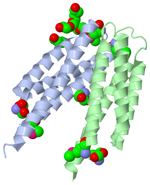
Asymmetric Unit (4, 10)
No.NameCountTypeFull Name
1MSE4Mod. Amino AcidSELENOMETHIONINE
2PEG2Ligand/IonDI(HYDROXYETHYL)ETHER
3PG41Ligand/IonTETRAETHYLENE GLYCOL
4TRS3Ligand/Ion2-AMINO-2-HYDROXYMETHYL-PROPANE-1,3-DIOL
Biological Unit 1 (4, 7)
No.NameCountTypeFull Name
1MSE2Mod. Amino AcidSELENOMETHIONINE
2PEG2Ligand/IonDI(HYDROXYETHYL)ETHER
3PG41Ligand/IonTETRAETHYLENE GLYCOL
4TRS2Ligand/Ion2-AMINO-2-HYDROXYMETHYL-PROPANE-1,3-DIOL
Biological Unit 2 (2, 3)
No.NameCountTypeFull Name
1MSE2Mod. Amino AcidSELENOMETHIONINE
2PEG-1Ligand/IonDI(HYDROXYETHYL)ETHER
3PG4-1Ligand/IonTETRAETHYLENE GLYCOL
4TRS1Ligand/Ion2-AMINO-2-HYDROXYMETHYL-PROPANE-1,3-DIOL

(-) Sites  (6, 6)

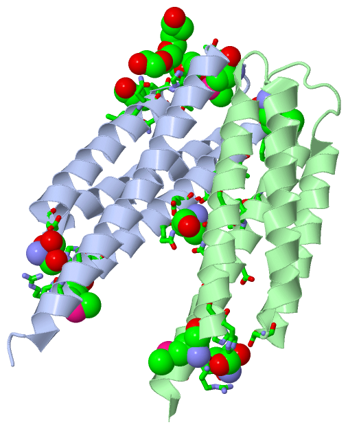
Asymmetric Unit (6, 6)
No.NameEvidenceResiduesDescription
1AC1SOFTWAREVAL A:68 , ARG A:72 , GLN A:116 , ALA A:119 , PEG A:205 , ASN B:140 , LEU B:144BINDING SITE FOR RESIDUE PG4 A 201
2AC2SOFTWAREARG A:45 , HIS A:48 , HIS A:49 , ASP A:100 , GLY A:156 , ALA A:160BINDING SITE FOR RESIDUE TRS A 202
3AC3SOFTWAREGLN A:57 , GLU A:60 , GLU B:60 , GLN B:147 , GLN B:150BINDING SITE FOR RESIDUE TRS A 203
4AC4SOFTWARELEU A:115 , GLN A:118 , ALA A:119 , LEU B:144 , GLN B:147 , ASP B:148BINDING SITE FOR RESIDUE PEG A 204
5AC5SOFTWAREGLN A:61 , ASP A:65 , ALA A:119 , HIS A:120 , THR A:121 , PRO A:122 , MSE A:129 , ALA A:130 , PG4 A:201 , ARG B:143BINDING SITE FOR RESIDUE PEG A 205
6AC6SOFTWAREARG B:45 , HIS B:48 , HIS B:49 , ASP B:100 , GLY B:156 , GLU B:159BINDING SITE FOR RESIDUE TRS B 201

(-) SS Bonds  (0, 0)

(no "SS Bond" information available for 4LLC)

(-) Cis Peptide Bonds  (0, 0)

(no "Cis Peptide Bond" information available for 4LLC)

 Sequence-Structure Mapping

(-) SAPs(SNPs)/Variants  (0, 0)

(no "SAP(SNP)/Variant" information available for 4LLC)

(-) PROSITE Motifs  (0, 0)

(no "PROSITE Motif" information available for 4LLC)

(-) Exons   (0, 0)

(no "Exon" information available for 4LLC)

(-) Sequences/Alignments

Asymmetric Unit
   Reformat: Number of residues per line =  ('0' or empty: single-line sequence representation)
  Number of residues per labelling interval =   
  UniProt sequence: complete  aligned part    
   Show mapping: SCOP domains CATH domains Pfam domains Secondary structure (by author)
SAPs(SNPs) PROSITE motifs Exons
(details for a mapped element are shown in a popup box when the mouse pointer rests over it)
Chain A from PDB  Type:PROTEIN  Length:124
                                                                                                                                                            
               SCOP domains ---------------------------------------------------------------------------------------------------------------------------- SCOP domains
               CATH domains ---------------------------------------------------------------------------------------------------------------------------- CATH domains
               Pfam domains ---------------------------------------------------------------------------------------------------------------------------- Pfam domains
         Sec.struct. author .hhhhhhhhhhhhhhhhhhhhhhhhhhh...hhhhhhhhhhhhhhhhhhhhhhh.hhhhhhhhhhhhhhhhhhhh...........hhhhhhhhhhhhhhhhhhhhhhhhhhhhhhhhhhh... Sec.struct. author
                 SAPs(SNPs) ---------------------------------------------------------------------------------------------------------------------------- SAPs(SNPs)
                    PROSITE ---------------------------------------------------------------------------------------------------------------------------- PROSITE
                 Transcript ---------------------------------------------------------------------------------------------------------------------------- Transcript
                 4llc A  45 RmSHHYATIEVSQQLEQLLGDQLVILLRETPDGQALERSQNDFRRVLEQGRANTVDSAEQAALDGVRDAYLQLQAHTPALLEAPmADNDGFSEAFNGLRLRLQDLQQLALAGISEAETSARHRA 168
                             |      54        64        74        84        94       104       114       124    |  134       144       154       164    
                             |                                                                                129-MSE                                   
                            46-MSE                                                                                                                      

Chain B from PDB  Type:PROTEIN  Length:120
                                                                                                                                                        
               SCOP domains ------------------------------------------------------------------------------------------------------------------------ SCOP domains
               CATH domains ------------------------------------------------------------------------------------------------------------------------ CATH domains
               Pfam domains ------------------------------------------------------------------------------------------------------------------------ Pfam domains
         Sec.struct. author .hhhhhhhhhhhhhhhhhhhhhhhhhhh...hhhhhhhhhhhhhhhhhhhhhhh.hhhhhhhhhhhhhhhhhhhhh...........hhhhhhhhhhhhhhhhhhhhhhhhhhhhhhhhh Sec.struct. author
                 SAPs(SNPs) ------------------------------------------------------------------------------------------------------------------------ SAPs(SNPs)
                    PROSITE ------------------------------------------------------------------------------------------------------------------------ PROSITE
                 Transcript ------------------------------------------------------------------------------------------------------------------------ Transcript
                 4llc B  45 RmSHHYATIEVSQQLEQLLGDQLVILLRETPDGQALERSQNDFRRVLEQGRANTVDSAEQAALDGVRDAYLQLQAHTPALLEAPmADNDGFSEAFNGLRLRLQDLQQLALAGISEAETSA 164
                             |      54        64        74        84        94       104       114       124    |  134       144       154       164
                            46-MSE                                                                            129-MSE                               

   Legend:   → Mismatch (orange background)
  - → Gap (green background, '-', border residues have a numbering label)
    → Modified Residue (blue background, lower-case, 'x' indicates undefined single-letter code, labelled with number + name)
  x → Chemical Group (purple background, 'x', labelled with number + name, e.g. ACE or NH2)
  extra numbering lines below/above indicate numbering irregularities and modified residue names etc., number ends below/above '|'

 Classification and Annotation

(-) SCOP Domains  (0, 0)

(no "SCOP Domain" information available for 4LLC)

(-) CATH Domains  (0, 0)

(no "CATH Domain" information available for 4LLC)

(-) Pfam Domains  (0, 0)

(no "Pfam Domain" information available for 4LLC)

(-) Gene Ontology  (16, 16)

Asymmetric Unit(hide GO term definitions)

 Visualization

(-) Interactive Views

Asymmetric Unit
  Complete Structure
    Jena3D(integrated viewing of ligand, site, SAP, PROSITE, SCOP information)
    WebMol | AstexViewer[tm]@PDBe
(Java Applets, require no local installation except for Java; loading may be slow)
    STRAP
(Java WebStart application, automatic local installation, requires Java; full application with system access!)
    RasMol
(require local installation)
    Molscript (VRML)
(requires installation of a VRML viewer; select preferred view via VRML and generate a mono or stereo PDF format file)
 
  Ligands, Modified Residues, Ions
    MSE  [ RasMol | Jena3D ]  +environment [ RasMol | Jena3D ]
    PEG  [ RasMol | Jena3D ]  +environment [ RasMol | Jena3D ]
    PG4  [ RasMol | Jena3D ]  +environment [ RasMol | Jena3D ]
    TRS  [ RasMol | Jena3D ]  +environment [ RasMol | Jena3D ]
 
  Sites
    AC1  [ RasMol ]  +environment [ RasMol ]
    AC2  [ RasMol ]  +environment [ RasMol ]
    AC3  [ RasMol ]  +environment [ RasMol ]
    AC4  [ RasMol ]  +environment [ RasMol ]
    AC5  [ RasMol ]  +environment [ RasMol ]
    AC6  [ RasMol ]  +environment [ RasMol ]
 
  Cis Peptide Bonds
(no "Cis Peptide Bonds" information available for 4llc)
 
Biological Units
  Complete Structure
    Biological Unit 1  [ Jena3D ]
    Biological Unit 2  [ Jena3D ]

(-) Still Images

Jmol
  protein: cartoon or spacefill or dots and stick; nucleic acid: cartoon and stick; ligands: spacefill; active site: stick
Molscript
  protein, nucleic acid: cartoon; ligands: spacefill; active site: ball and stick

 Databases and Analysis Tools

(-) Databases

Access by PDB/NDB ID
  4llc
    Family and Domain InformationProDom | SYSTERS
    General Structural InformationGlycoscienceDB | MMDB | NDB | OCA | PDB | PDBe | PDBj | PDBsum | PDBWiki | PQS | PROTEOPEDIA
    Orientation in MembranesOPM
    Protein SurfaceSURFACE
    Secondary StructureDSSP (structure derived) | HSSP (homology derived)
    Structural GenomicsGeneCensus
    Structural NeighboursCE | VAST
    Structure ClassificationCATH | Dali | SCOP
    Validation and Original DataBMRB Data View | BMRB Restraints Grid | EDS | PROCHECK | RECOORD | WHAT_CHECK
 
Access by UniProt ID/Accession number
  Q9HT87_PSEAE | Q9HT87
    Comparative Protein Structure ModelsModBase
    Genomic InformationEnsembl
    Protein-protein InteractionDIP
    Sequence, Family and Domain InformationInterPro | Pfam | SMART | UniProtKB/TrEMBL
 
Access by Enzyme Classificator   (EC Number)
  (no 'Enzyme Classificator' available)
    General Enzyme InformationBRENDA | EC-PDB | Enzyme | IntEnz
    PathwayKEGG | MetaCyc
 
Access by Disease Identifier   (MIM ID)
  (no 'MIM ID' available)
    Disease InformationOMIM
 
Access by GenAge ID
  (no 'GenAge ID' available)
    Age Related InformationGenAge

(-) Analysis Tools

Access by PDB/NDB ID
    Domain InformationXDom
    Interatomic Contacts of Structural UnitsCSU
    Ligand-protein ContactsLPC
    Protein CavitiescastP
    Sequence and Secondary StructurePDBCartoon
    Structure AlignmentSTRAP(Java WebStart application, automatic local installation, requires Java; full application with system access!)
    Structure and Sequence BrowserSTING
 
Access by UniProt ID/Accession number
  Q9HT87_PSEAE | Q9HT87
    Protein Disorder PredictionDisEMBL | FoldIndex | GLOBPLOT (for more information see DisProt)

 Related Entries

(-) Entries Sharing at Least One Protein Chain (UniProt ID)

UniProtKB/Swiss-Prot
        Q9HT87_PSEAE | Q9HT873kkb
UniProtKB/TrEMBL
        Q9HT87_PSEAE | Q9HT873l34 3n24 4lle

(-) Related Entries Specified in the PDB File

3kkb PO4-BOUND
3l34 LIGAND-FREE
4lle R60L RELATED ID: MCSG-APC105591 RELATED DB: TARGETTRACK