Show PDB file:   
         Plain Text   HTML   (compressed file size)
QuickSearch:   
by PDB,NDB,UniProt,PROSITE Code or Search Term(s)  
(-)Asymmetric Unit
(-)Asym. Unit - sites
(-)Biological Unit 1
(-)Biol. Unit 1 - sites
(-)Biological Unit 2
(-)Biological Unit 3
collapse expand < >
Image Asymmetric Unit
Asymmetric Unit  (Jmol Viewer)
Image Asym. Unit - sites
Asym. Unit - sites  (Jmol Viewer)
Image Biological Unit 1
Biological Unit 1  (Jmol Viewer)
Image Biol. Unit 1 - sites
Biol. Unit 1 - sites  (Jmol Viewer)
Image Biological Unit 2
Biological Unit 2  (Jmol Viewer)
Image Biological Unit 3
Biological Unit 3  (Jmol Viewer)

(-) Description

Title :  CRYSTAL STRUCTURE OF ATBAG3
 
Authors :  Y. Shen, S. Fang
Date :  07 Nov 12  (Deposition) - 29 May 13  (Release) - 19 Jun 13  (Revision)
Method :  X-RAY DIFFRACTION
Resolution :  2.00
Chains :  Asym. Unit :  A,B
Biol. Unit 1:  A  (1x)
Biol. Unit 2:  B  (1x)
Biol. Unit 3:  A,B  (1x)
Keywords :  Three Helix Bundle, Co-Chaperone, Apoptosis (Keyword Search: [Gene Ontology, PubMed, Web (Google))
 
Reference :  S. Fang, L. Li, B. Cui, S. Men, Y. Shen, X. Yang
Structural Insight Into Plant Programmed Cell Death Mediate By Bag Proteins In Arabidopsis Thaliana.
Acta Crystallogr. , Sect. D V. 69 934 2013
PubMed-ID: 23695238  |  Reference-DOI: 10.1107/S0907444913003624

(-) Compounds

Molecule 1 - BAG FAMILY MOLECULAR CHAPERONE REGULATOR 3
    ChainsA, B
    EngineeredYES
    Expression SystemESCHERICHIA COLI
    Expression System Taxid562
    FragmentBAG DOMAIN (UNP RESIDUES 135-220)
    GeneAT5G07220, BAG3, T28J14_160
    Organism CommonMOUSE-EAR CRESS
    Organism ScientificARABIDOPSIS THALIANA
    Organism Taxid3702
    SynonymBCL-2-ASSOCIATED ATHANOGENE 3

 Structural Features

(-) Chains, Units

  12
Asymmetric Unit AB
Biological Unit 1 (1x)A 
Biological Unit 2 (1x) B
Biological Unit 3 (1x)AB

Summary Information (see also Sequences/Alignments below)

(-) Ligands, Modified Residues, Ions  (1, 2)

Asymmetric Unit (1, 2)
No.NameCountTypeFull Name
1SO42Ligand/IonSULFATE ION
Biological Unit 1 (1, 1)
No.NameCountTypeFull Name
1SO41Ligand/IonSULFATE ION
Biological Unit 2 (1, 1)
No.NameCountTypeFull Name
1SO41Ligand/IonSULFATE ION
Biological Unit 3 (1, 2)
No.NameCountTypeFull Name
1SO42Ligand/IonSULFATE ION

(-) Sites  (2, 2)

Asymmetric Unit (2, 2)
No.NameEvidenceResiduesDescription
1AC1SOFTWARETHR A:158 , ASN A:161 , HOH A:473BINDING SITE FOR RESIDUE SO4 A 301
2AC2SOFTWARELYS A:207 , GLY B:151 , GLN B:152BINDING SITE FOR RESIDUE SO4 B 301

(-) SS Bonds  (0, 0)

(no "SS Bond" information available for 4HWF)

(-) Cis Peptide Bonds  (0, 0)

(no "Cis Peptide Bond" information available for 4HWF)

 Sequence-Structure Mapping

(-) SAPs(SNPs)/Variants  (0, 0)

(no "SAP(SNP)/Variant" information available for 4HWF)

(-) PROSITE Motifs  (0, 0)

(no "PROSITE Motif" information available for 4HWF)

(-) Exons   (0, 0)

(no "Exon" information available for 4HWF)

(-) Sequences/Alignments

Asymmetric Unit
   Reformat: Number of residues per line =  ('0' or empty: single-line sequence representation)
  Number of residues per labelling interval =   
  UniProt sequence: complete  aligned part    
   Show mapping: SCOP domains CATH domains Pfam domains Secondary structure (by author)
SAPs(SNPs) PROSITE motifs Exons
(details for a mapped element are shown in a popup box when the mouse pointer rests over it)
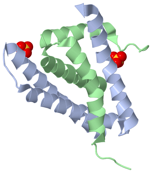
Chain A from PDB  Type:PROTEIN  Length:87
                                                                                                                       
               SCOP domains --------------------------------------------------------------------------------------- SCOP domains
               CATH domains --------------------------------------------------------------------------------------- CATH domains
               Pfam domains --------------------------------------------------------------------------------------- Pfam domains
         Sec.struct. author hhhhhhhhhhhhhhhhhhhhhhhhhhhhh....hhhhhhhhhhhhhhhhhhhhhhhh..hhhhhhhhhhhhhhhhhhhhhhhhhhh. Sec.struct. author
                 SAPs(SNPs) --------------------------------------------------------------------------------------- SAPs(SNPs)
                    PROSITE --------------------------------------------------------------------------------------- PROSITE
                 Transcript --------------------------------------------------------------------------------------- Transcript
                 4hwf A 134 SASKSISDISFEVDRLAGQVSAFETVINKGGKVEEKSLVNLIEMLMNQLLRLDAIIADGDVKLMRKMQVQRVQKYVEALDLLKVKNS 220
                                   143       153       163       173       183       193       203       213       

Chain B from PDB  Type:PROTEIN  Length:75
                                                                                                           
               SCOP domains --------------------------------------------------------------------------- SCOP domains
               CATH domains --------------------------------------------------------------------------- CATH domains
               Pfam domains --------------------------------------------------------------------------- Pfam domains
         Sec.struct. author .hhhhhhhhhhhhhhhh........hhhhhhhhhhhhhhhhhhhhhh.hhhhhhhhhhhhhhhhhhhhh...... Sec.struct. author
                 SAPs(SNPs) --------------------------------------------------------------------------- SAPs(SNPs)
                    PROSITE --------------------------------------------------------------------------- PROSITE
                 Transcript --------------------------------------------------------------------------- Transcript
                 4hwf B 134 SASKSISDISFEVDRLAGQVSAFEKSLVNLIEMLMNQLLRLDAIIADGDVKLMRKMQVQRVQKYVEALDLLKVKN 219
                                   143       153  ||   174       184       194       204       214     
                                                156|                                                   
                                                 168                                                   

   Legend:   → Mismatch (orange background)
  - → Gap (green background, '-', border residues have a numbering label)
    → Modified Residue (blue background, lower-case, 'x' indicates undefined single-letter code, labelled with number + name)
  x → Chemical Group (purple background, 'x', labelled with number + name, e.g. ACE or NH2)
  extra numbering lines below/above indicate numbering irregularities and modified residue names etc., number ends below/above '|'

 Classification and Annotation

(-) SCOP Domains  (0, 0)

(no "SCOP Domain" information available for 4HWF)

(-) CATH Domains  (0, 0)

(no "CATH Domain" information available for 4HWF)

(-) Pfam Domains  (0, 0)

(no "Pfam Domain" information available for 4HWF)

(-) Gene Ontology  (1, 1)

Asymmetric Unit(hide GO term definitions)

 Visualization

(-) Interactive Views

Asymmetric Unit
  Complete Structure
    Jena3D(integrated viewing of ligand, site, SAP, PROSITE, SCOP information)
    WebMol | AstexViewer[tm]@PDBe
(Java Applets, require no local installation except for Java; loading may be slow)
    STRAP
(Java WebStart application, automatic local installation, requires Java; full application with system access!)
    RasMol
(require local installation)
    Molscript (VRML)
(requires installation of a VRML viewer; select preferred view via VRML and generate a mono or stereo PDF format file)
 
  Ligands, Modified Residues, Ions
    SO4  [ RasMol | Jena3D ]  +environment [ RasMol | Jena3D ]
 
  Sites
    AC1  [ RasMol ]  +environment [ RasMol ]
    AC2  [ RasMol ]  +environment [ RasMol ]
 
  Cis Peptide Bonds
(no "Cis Peptide Bonds" information available for 4hwf)
 
Biological Units
  Complete Structure
    Biological Unit 1  [ Jena3D ]
    Biological Unit 2  [ Jena3D ]
    Biological Unit 3  [ Jena3D ]

(-) Still Images

Jmol
  protein: cartoon or spacefill or dots and stick; nucleic acid: cartoon and stick; ligands: spacefill; active site: stick
Molscript
  protein, nucleic acid: cartoon; ligands: spacefill; active site: ball and stick

 Databases and Analysis Tools

(-) Databases

Access by PDB/NDB ID
  4hwf
    Family and Domain InformationProDom | SYSTERS
    General Structural InformationGlycoscienceDB | MMDB | NDB | OCA | PDB | PDBe | PDBj | PDBsum | PDBWiki | PQS | PROTEOPEDIA
    Orientation in MembranesOPM
    Protein SurfaceSURFACE
    Secondary StructureDSSP (structure derived) | HSSP (homology derived)
    Structural GenomicsGeneCensus
    Structural NeighboursCE | VAST
    Structure ClassificationCATH | Dali | SCOP
    Validation and Original DataBMRB Data View | BMRB Restraints Grid | EDS | PROCHECK | RECOORD | WHAT_CHECK
 
Access by UniProt ID/Accession number
  BAG3_ARATH | Q9LYP4
    Comparative Protein Structure ModelsModBase
    Genomic InformationEnsembl
    Protein-protein InteractionDIP
    Sequence, Family and Domain InformationInterPro | Pfam | SMART | UniProtKB/SwissProt
 
Access by Enzyme Classificator   (EC Number)
  (no 'Enzyme Classificator' available)
    General Enzyme InformationBRENDA | EC-PDB | Enzyme | IntEnz
    PathwayKEGG | MetaCyc
 
Access by Disease Identifier   (MIM ID)
  (no 'MIM ID' available)
    Disease InformationOMIM
 
Access by GenAge ID
  (no 'GenAge ID' available)
    Age Related InformationGenAge

(-) Analysis Tools

Access by PDB/NDB ID
    Domain InformationXDom
    Interatomic Contacts of Structural UnitsCSU
    Ligand-protein ContactsLPC
    Protein CavitiescastP
    Sequence and Secondary StructurePDBCartoon
    Structure AlignmentSTRAP(Java WebStart application, automatic local installation, requires Java; full application with system access!)
    Structure and Sequence BrowserSTING
 
Access by UniProt ID/Accession number
  BAG3_ARATH | Q9LYP4
    Protein Disorder PredictionDisEMBL | FoldIndex | GLOBPLOT (for more information see DisProt)

 Related Entries

(-) Entries Sharing at Least One Protein Chain (UniProt ID)

(no "Entries Sharing at Least One Protein Chain" available for 4HWF)

(-) Related Entries Specified in the PDB File

4hwc 4hwd 4hwh 4hwi