Show PDB file:   
         Plain Text   HTML   (compressed file size)
QuickSearch:   
by PDB,NDB,UniProt,PROSITE Code or Search Term(s)  
(-)Asymmetric Unit
(-)Asym. Unit - sites
(-)Biological Unit 1
(-)Biol. Unit 1 - sites
(-)Biological Unit 2
collapse expand < >
Image Asymmetric Unit
Asymmetric Unit  (Jmol Viewer)
Image Asym. Unit - sites
Asym. Unit - sites  (Jmol Viewer)
Image Biological Unit 1
Biological Unit 1  (Jmol Viewer)
Image Biol. Unit 1 - sites
Biol. Unit 1 - sites  (Jmol Viewer)
Image Biological Unit 2
Biological Unit 2  (Jmol Viewer)

(-) Description

Title :  CRYSTAL STRUCTURE OF MAGNESIUM-LOADED PLASMODIUM VIVAX TRAP PROTEIN
 
Authors :  G. Song, A. C. Koksal, C. Lu, T. A. Springer
Date :  25 Oct 12  (Deposition) - 26 Dec 12  (Release) - 09 Jan 13  (Revision)
Method :  X-RAY DIFFRACTION
Resolution :  2.24
Chains :  Asym. Unit :  A,B
Biol. Unit 1:  A  (1x)
Biol. Unit 2:  B  (1x)
Keywords :  Malaria, Parasite Motility, Vwa Domain, Tsr Domain, Receptor On Sporozoite, Vaccine Target, Sporozoite Surface, Cell Adhesion (Keyword Search: [Gene Ontology, PubMed, Web (Google))
 
Reference :  G. Song, A. C. Koksal, C. Lu, T. A. Springer
Shape Change In The Receptor For Gliding Motility In Plasmodium Sporozoites.
Proc. Natl. Acad. Sci. Usa V. 109 21420 2012
PubMed-ID: 23236185  |  Reference-DOI: 10.1073/PNAS.1218581109

(-) Compounds

Molecule 1 - SPOROZOITE SURFACE PROTEIN 2
    ChainsA, B
    EngineeredYES
    Expression SystemHOMO SAPIENS
    Expression System Cell LineHEK293T
    Expression System CommonHUMAN
    Expression System Taxid9606
    FragmentADHESIVE DOMAINS (UNP RESIDUES 25-283)
    GeneSSP2
    MutationYES
    Organism ScientificPLASMODIUM VIVAX
    Organism Taxid5855
    StrainSAL I
    SynonymTHROMBOSPONDIN REPEAT ANONYMOUS PROTEIN, TRAP

 Structural Features

(-) Chains, Units

  12
Asymmetric Unit AB
Biological Unit 1 (1x)A 
Biological Unit 2 (1x) B

Summary Information (see also Sequences/Alignments below)

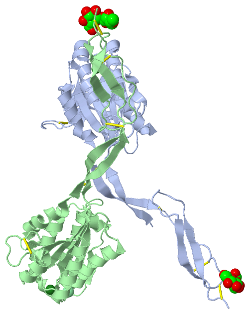
(-) Ligands, Modified Residues, Ions  (4, 8)

Asymmetric Unit (4, 8)
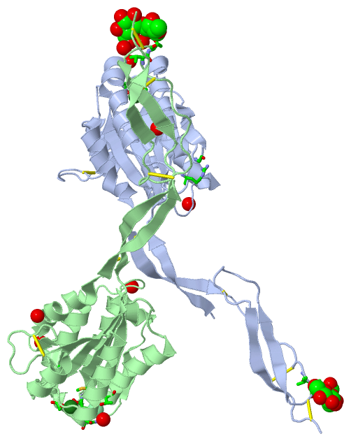
No.NameCountTypeFull Name
1BGC2Ligand/IonBETA-D-GLUCOSE
2CL2Ligand/IonCHLORIDE ION
3FUC2Ligand/IonALPHA-L-FUCOSE
4MG2Ligand/IonMAGNESIUM ION
Biological Unit 1 (2, 2)
No.NameCountTypeFull Name
1BGC1Ligand/IonBETA-D-GLUCOSE
2CL-1Ligand/IonCHLORIDE ION
3FUC1Ligand/IonALPHA-L-FUCOSE
4MG-1Ligand/IonMAGNESIUM ION
Biological Unit 2 (2, 2)
No.NameCountTypeFull Name
1BGC1Ligand/IonBETA-D-GLUCOSE
2CL-1Ligand/IonCHLORIDE ION
3FUC1Ligand/IonALPHA-L-FUCOSE
4MG-1Ligand/IonMAGNESIUM ION

(-) Sites  (6, 6)

Asymmetric Unit (6, 6)
No.NameEvidenceResiduesDescription
1AC1SOFTWARESER A:52 , SER A:54 , THR A:127 , HOH A:401 , HOH A:402 , HOH A:403BINDING SITE FOR RESIDUE MG A 301
2AC2SOFTWARETHR A:127 , MET A:129 , THR A:130 , ASN A:162 , SER A:163BINDING SITE FOR RESIDUE CL A 304
3AC3SOFTWARESER B:52 , SER B:54 , THR B:127 , HOH B:401 , HOH B:402BINDING SITE FOR RESIDUE MG B 301
4AC4SOFTWARETHR B:127 , MET B:129 , THR B:130 , ASN B:162 , SER B:163 , HOH B:442BINDING SITE FOR RESIDUE CL B 304
5AC5SOFTWAREVAL A:251 , THR A:252 , HOH A:605 , HOH B:639BINDING SITE FOR LINKED RESIDUES A 302 to 303
6AC6SOFTWAREARG A:79 , GLY A:100 , HOH A:532 , ILE B:61 , VAL B:251 , THR B:252 , HOH B:410 , HOH B:417 , HOH B:545 , HOH B:547 , HOH B:564BINDING SITE FOR LINKED RESIDUES B 302 to 303

(-) SS Bonds  (10, 10)

Asymmetric Unit
No.Residues
1A:39 -A:231
2A:201 -A:208
3A:240 -A:269
4A:249 -A:277
5A:253 -A:282
6B:39 -B:231
7B:201 -B:208
8B:240 -B:269
9B:249 -B:277
10B:253 -B:282

(-) Cis Peptide Bonds  (0, 0)

(no "Cis Peptide Bond" information available for 4HQL)

 Sequence-Structure Mapping

(-) SAPs(SNPs)/Variants  (0, 0)

(no "SAP(SNP)/Variant" information available for 4HQL)

(-) PROSITE Motifs  (0, 0)

(no "PROSITE Motif" information available for 4HQL)

(-) Exons   (0, 0)

(no "Exon" information available for 4HQL)

(-) Sequences/Alignments

Asymmetric Unit
   Reformat: Number of residues per line =  ('0' or empty: single-line sequence representation)
  Number of residues per labelling interval =   
  UniProt sequence: complete  aligned part    
   Show mapping: SCOP domains CATH domains Pfam domains Secondary structure (by author)
SAPs(SNPs) PROSITE motifs Exons
(details for a mapped element are shown in a popup box when the mouse pointer rests over it)
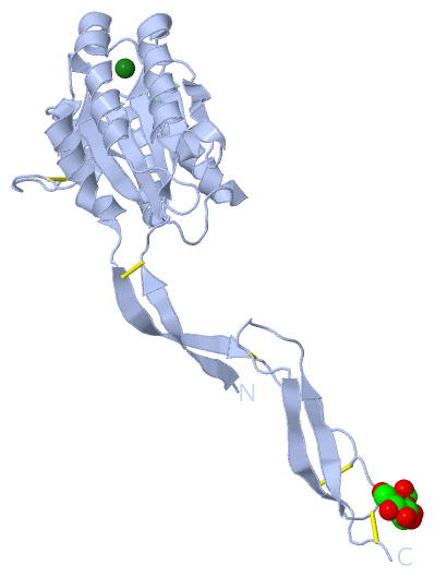
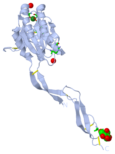
Chain A from PDB  Type:PROTEIN  Length:254
                                                                                                                                                                                                                                                                                              
               SCOP domains -------------------------------------------------------------------------------------------------------------------------------------------------------------------------------------------------------------------------------------------------------------- SCOP domains
               CATH domains -------------------------------------------------------------------------------------------------------------------------------------------------------------------------------------------------------------------------------------------------------------- CATH domains
               Pfam domains -------------------------------------------------------------------------------------------------------------------------------------------------------------------------------------------------------------------------------------------------------------- Pfam domains
         Sec.struct. author .eeeeeeee....eeeeeeee.....hhhhhhhhhhhhhhhhhhh.......eeeeeeee...eeeee...hhhhhhhhhhhhhhhhhhhhh......hhhhhhhhhhhhhhh.......eeeeeeee.....hhhhhhhhhhhhhh...eeeeee.....hhhhhhhhhh..........ee..hhhhhhhhhhhhhh..eeeeeeeee...............eeeeeee..........eeeeeee..... Sec.struct. author
                 SAPs(SNPs) -------------------------------------------------------------------------------------------------------------------------------------------------------------------------------------------------------------------------------------------------------------- SAPs(SNPs)
                    PROSITE -------------------------------------------------------------------------------------------------------------------------------------------------------------------------------------------------------------------------------------------------------------- PROSITE
                 Transcript -------------------------------------------------------------------------------------------------------------------------------------------------------------------------------------------------------------------------------------------------------------- Transcript
                 4hql A  30 DEVKYSEEVCNEQVDLYLLVDGSGSIGYPNWITKVIPMLNGLINSLSLSRDTINLYMNLFGSYTTELIRLGSGQSIDKRQALSKVTELRKTYTPYGTTSMTAALDEVQKHLNDRVNREKAIQLVILMTDGVPNSKYRALEVANKLKQRNVRLAVIGIGQGINHQFNRLIAGCRPREPNCKFYSYADWNEAVALIKPFIAKVCTEVERVANCGPWDPWTACSVTCGRGTHSRSRPSLHEKCTTHMVSECEEGECP 283
                                    39        49        59        69        79        89        99       109       119       129       139       149       159       169       179       189       199       209       219       229       239       249       259       269       279    

Chain B from PDB  Type:PROTEIN  Length:256
                                                                                                                                                                                                                                                                                                
               SCOP domains ---------------------------------------------------------------------------------------------------------------------------------------------------------------------------------------------------------------------------------------------------------------- SCOP domains
               CATH domains ---------------------------------------------------------------------------------------------------------------------------------------------------------------------------------------------------------------------------------------------------------------- CATH domains
               Pfam domains ---------------------------------------------------------------------------------------------------------------------------------------------------------------------------------------------------------------------------------------------------------------- Pfam domains
         Sec.struct. author ...eeeeeeeeee..eeeeeeee.hhhhhhhhhhhhhhhhhhhhhhh.......eeeeeeee...eeeee...hhhhhhhhhhhhhhhhhhhhh......hhhhhhhhhhhhhhh.......eeeeeeee.....hhhhhhhhhhhhhhh..eeeeee.....hhhhhhhhhh..........ee..hhhhhhhhhhhhhh.eeeeeeeeee...............eeeeeee..........eeeeeee..... Sec.struct. author
                 SAPs(SNPs) ---------------------------------------------------------------------------------------------------------------------------------------------------------------------------------------------------------------------------------------------------------------- SAPs(SNPs)
                    PROSITE ---------------------------------------------------------------------------------------------------------------------------------------------------------------------------------------------------------------------------------------------------------------- PROSITE
                 Transcript ---------------------------------------------------------------------------------------------------------------------------------------------------------------------------------------------------------------------------------------------------------------- Transcript
                 4hql B  28 VVDEVKYSEEVCNEQVDLYLLVDGSGSIGYPNWITKVIPMLNGLINSLSLSRDTINLYMNLFGSYTTELIRLGSGQSIDKRQALSKVTELRKTYTPYGTTSMTAALDEVQKHLNDRVNREKAIQLVILMTDGVPNSKYRALEVANKLKQRNVRLAVIGIGQGINHQFNRLIAGCRPREPNCKFYSYADWNEAVALIKPFIAKVCTEVERVANCGPWDPWTACSVTCGRGTHSRSRPSLHEKCTTHMVSECEEGECP 283
                                    37        47        57        67        77        87        97       107       117       127       137       147       157       167       177       187       197       207       217       227       237       247       257       267       277      

   Legend:   → Mismatch (orange background)
  - → Gap (green background, '-', border residues have a numbering label)
    → Modified Residue (blue background, lower-case, 'x' indicates undefined single-letter code, labelled with number + name)
  x → Chemical Group (purple background, 'x', labelled with number + name, e.g. ACE or NH2)
  extra numbering lines below/above indicate numbering irregularities and modified residue names etc., number ends below/above '|'

 Classification and Annotation

(-) SCOP Domains  (0, 0)

(no "SCOP Domain" information available for 4HQL)

(-) CATH Domains  (0, 0)

(no "CATH Domain" information available for 4HQL)

(-) Pfam Domains  (0, 0)

(no "Pfam Domain" information available for 4HQL)

(-) Gene Ontology  (3, 3)

Asymmetric Unit(hide GO term definitions)

 Visualization

(-) Interactive Views

Asymmetric Unit
  Complete Structure
    Jena3D(integrated viewing of ligand, site, SAP, PROSITE, SCOP information)
    WebMol | AstexViewer[tm]@PDBe
(Java Applets, require no local installation except for Java; loading may be slow)
    STRAP
(Java WebStart application, automatic local installation, requires Java; full application with system access!)
    RasMol
(require local installation)
    Molscript (VRML)
(requires installation of a VRML viewer; select preferred view via VRML and generate a mono or stereo PDF format file)
 
  Ligands, Modified Residues, Ions
    BGC  [ RasMol | Jena3D ]  +environment [ RasMol | Jena3D ]
    CL  [ RasMol | Jena3D ]  +environment [ RasMol | Jena3D ]
    FUC  [ RasMol | Jena3D ]  +environment [ RasMol | Jena3D ]
    MG  [ RasMol | Jena3D ]  +environment [ RasMol | Jena3D ]
 
  Sites
    AC1  [ RasMol ]  +environment [ RasMol ]
    AC2  [ RasMol ]  +environment [ RasMol ]
    AC3  [ RasMol ]  +environment [ RasMol ]
    AC4  [ RasMol ]  +environment [ RasMol ]
    AC5  [ RasMol ]  +environment [ RasMol ]
    AC6  [ RasMol ]  +environment [ RasMol ]
 
  Cis Peptide Bonds
(no "Cis Peptide Bonds" information available for 4hql)
 
Biological Units
  Complete Structure
    Biological Unit 1  [ Jena3D ]
    Biological Unit 2  [ Jena3D ]

(-) Still Images

Jmol
  protein: cartoon or spacefill or dots and stick; nucleic acid: cartoon and stick; ligands: spacefill; active site: stick
Molscript
  protein, nucleic acid: cartoon; ligands: spacefill; active site: ball and stick

 Databases and Analysis Tools

(-) Databases

Access by PDB/NDB ID
  4hql
    Family and Domain InformationProDom | SYSTERS
    General Structural InformationGlycoscienceDB | MMDB | NDB | OCA | PDB | PDBe | PDBj | PDBsum | PDBWiki | PQS | PROTEOPEDIA
    Orientation in MembranesOPM
    Protein SurfaceSURFACE
    Secondary StructureDSSP (structure derived) | HSSP (homology derived)
    Structural GenomicsGeneCensus
    Structural NeighboursCE | VAST
    Structure ClassificationCATH | Dali | SCOP
    Validation and Original DataBMRB Data View | BMRB Restraints Grid | EDS | PROCHECK | RECOORD | WHAT_CHECK
 
Access by UniProt ID/Accession number
  Q9TVF0_PLAVI | Q9TVF0
    Comparative Protein Structure ModelsModBase
    Genomic InformationEnsembl
    Protein-protein InteractionDIP
    Sequence, Family and Domain InformationInterPro | Pfam | SMART | UniProtKB/TrEMBL
 
Access by Enzyme Classificator   (EC Number)
  (no 'Enzyme Classificator' available)
    General Enzyme InformationBRENDA | EC-PDB | Enzyme | IntEnz
    PathwayKEGG | MetaCyc
 
Access by Disease Identifier   (MIM ID)
  (no 'MIM ID' available)
    Disease InformationOMIM
 
Access by GenAge ID
  (no 'GenAge ID' available)
    Age Related InformationGenAge

(-) Analysis Tools

Access by PDB/NDB ID
    Domain InformationXDom
    Interatomic Contacts of Structural UnitsCSU
    Ligand-protein ContactsLPC
    Protein CavitiescastP
    Sequence and Secondary StructurePDBCartoon
    Structure AlignmentSTRAP(Java WebStart application, automatic local installation, requires Java; full application with system access!)
    Structure and Sequence BrowserSTING
 
Access by UniProt ID/Accession number
  Q9TVF0_PLAVI | Q9TVF0
    Protein Disorder PredictionDisEMBL | FoldIndex | GLOBPLOT (for more information see DisProt)

 Related Entries

(-) Entries Sharing at Least One Protein Chain (UniProt ID)

UniProtKB/TrEMBL
        Q9TVF0_PLAVI | Q9TVF04hqn 4hqo

(-) Related Entries Specified in the PDB File

4hqf 4hqk 4hqn 4hqo