|
|
|
|
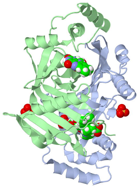
 Description
|
|
Compounds
|
||||||||||||||||||||||||||||||||||||||||||||||||||||
Chains, Units
Summary Information (see also Sequences/Alignments below) |
Ligands, Modified Residues, Ions (3, 5)| Asymmetric/Biological Unit (3, 5) |
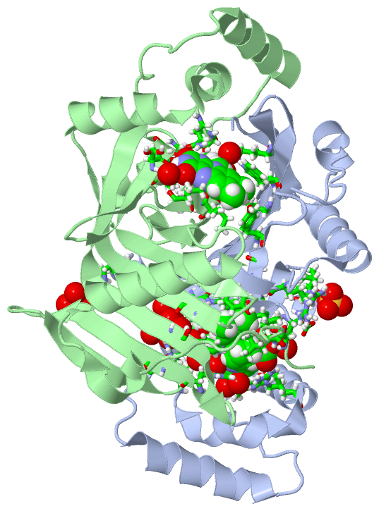
Sites (5, 5)
Asymmetric Unit (5, 5)
|
SS Bonds (0, 0)| (no "SS Bond" information available for 4HMU) |
Cis Peptide Bonds (1, 1)
Asymmetric/Biological Unit
|
||||||||
SAPs(SNPs)/Variants (0, 0)| (no "SAP(SNP)/Variant" information available for 4HMU) |
PROSITE Motifs (0, 0)| (no "PROSITE Motif" information available for 4HMU) |
Exons (0, 0)| (no "Exon" information available for 4HMU) |
Sequences/Alignments
Asymmetric/Biological Unit
Chain A from PDB Type:PROTEIN Length:202
SCOP domains d4hmua_ A: Pyridoxine 5'-phoshate oxidase (PNP oxidase) SCOP domains
CATH domains ---------------------------------------------------------------------------------------------------------------------------------------------------------------------------------------------------------- CATH domains
Pfam domains ---------------------------------------------------------------------------------------------------------------------------------------------------------------------------------------------------------- Pfam domains
SAPs(SNPs) ---------------------------------------------------------------------------------------------------------------------------------------------------------------------------------------------------------- SAPs(SNPs)
PROSITE ---------------------------------------------------------------------------------------------------------------------------------------------------------------------------------------------------------- PROSITE
Transcript ---------------------------------------------------------------------------------------------------------------------------------------------------------------------------------------------------------- Transcript
4hmu A 21 GTLDAPFPEYQTLPADPMSVLHNWLERARRVGIREPRALALATADSQGRPSTRIVVISEISDAGVVFSTHAGSQKGRELLHNPWASGVLYWRETSQQIILNGQAVRLPNAKADDAWLKRPYATHPMSSVSRQSEELQDVQAMRNAARQLAELQGPLPRPEGYCVFELRLESLEFWGNGQERLHERLRYDRSDTGWNVRRLQP 222
30 40 50 60 70 80 90 100 110 120 130 140 150 160 170 180 190 200 210 220
Chain B from PDB Type:PROTEIN Length:207
SCOP domains d4hmub_ B: Pyridoxine 5'-phoshate oxidase (PNP oxidase) SCOP domains
CATH domains --------------------------------------------------------------------------------------------------------------------------------------------------------------------------------------------------------------- CATH domains
Pfam domains --------------------------------------------------------------------------------------------------------------------------------------------------------------------------------------------------------------- Pfam domains
SAPs(SNPs) --------------------------------------------------------------------------------------------------------------------------------------------------------------------------------------------------------------- SAPs(SNPs)
PROSITE --------------------------------------------------------------------------------------------------------------------------------------------------------------------------------------------------------------- PROSITE
Transcript --------------------------------------------------------------------------------------------------------------------------------------------------------------------------------------------------------------- Transcript
4hmu B 16 SESLTGTLDAPFPEYQTLPADPMSVLHNWLERARRVGIREPRALALATADSQGRPSTRIVVISEISDAGVVFSTHAGSQKGRELLHNPWASGVLYWRETSQQIILNGQAVRLPNAKADDAWLKRPYATHPMSSVSRQSEELQDVQAMRNAARQLAELQGPLPRPEGYCVFELRLESLEFWGNGQERLHERLRYDRSDTGWNVRRLQP 222
25 35 45 55 65 75 85 95 105 115 125 135 145 155 165 175 185 195 205 215
|
||||||||||||||||||||
SCOP Domains (1, 2)
Asymmetric/Biological Unit
 |
CATH Domains (0, 0)| (no "CATH Domain" information available for 4HMU) |
Pfam Domains (0, 0)| (no "Pfam Domain" information available for 4HMU) |
Gene Ontology (10, 10)|
Asymmetric/Biological Unit(hide GO term definitions) |
Interactive Views
|
|||||||||||||||||||||||||||||||||||||||||||||||||||||||||||||||||||||||||||||||||||||||||||||||||||||||||||||||||||||||||||||||||||||||||||||||||||||||||||||||||
Still Images
|
||||||||||||||||
Databases
|
||||||||||||||||||||||||||||||||||||||||||||||||||||||||||||||||||||||||||||||||||||||||||||||||||||||||||||||||||||||||||||||||||||||||||||||||||||||||||||||||
Analysis Tools
|
|||||||||||||||||||||||||||||||||||||||||||||||||||||||||||||
Entries Sharing at Least One Protein Chain (UniProt ID)
Related Entries Specified in the PDB File
|
|