|
|
|
|
 Description
|
|
Compounds
|
||||||||||||||||||||||||||||||||||||||||||||||||||||
Chains, Units
Summary Information (see also Sequences/Alignments below) |
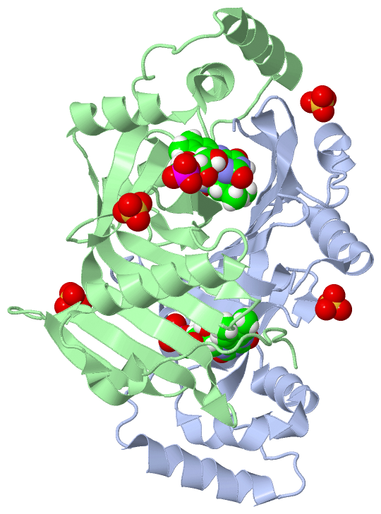
Ligands, Modified Residues, Ions (2, 7)| Asymmetric/Biological Unit (2, 7) |
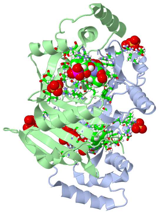
Sites (7, 7)
Asymmetric Unit (7, 7)
|
SS Bonds (0, 0)| (no "SS Bond" information available for 4HMS) |
Cis Peptide Bonds (1, 1)
Asymmetric/Biological Unit
|
||||||||
SAPs(SNPs)/Variants (0, 0)| (no "SAP(SNP)/Variant" information available for 4HMS) |
PROSITE Motifs (0, 0)| (no "PROSITE Motif" information available for 4HMS) |
Exons (0, 0)| (no "Exon" information available for 4HMS) |
Sequences/Alignments
Asymmetric/Biological Unit
Chain A from PDB Type:PROTEIN Length:202
SCOP domains d4hmsa_ A: Pyridoxine 5'-phoshate oxidase (PNP oxidase) SCOP domains
CATH domains ---------------------------------------------------------------------------------------------------------------------------------------------------------------------------------------------------------- CATH domains
Pfam domains ---------------------------------------------------------------------------------------------------------------------------------------------------------------------------------------------------------- Pfam domains
SAPs(SNPs) ---------------------------------------------------------------------------------------------------------------------------------------------------------------------------------------------------------- SAPs(SNPs)
PROSITE ---------------------------------------------------------------------------------------------------------------------------------------------------------------------------------------------------------- PROSITE
Transcript ---------------------------------------------------------------------------------------------------------------------------------------------------------------------------------------------------------- Transcript
4hms A 21 GTLDAPFPEYQTLPADPMSVLHNWLERARRVGIREPRALALATADSQGRPSTRIVVISEISDAGVVFSTHAGSQKGRELLHNPWASGVLYWRETSQQIILNGQAVRLPNAKADDAWLKRPYATHPMSSVSRQSEELQDVQAMRNAARQLAELQGPLPRPEGYCVFELRLESLEFWGNGQERLHERLRYDRSDTGWNVRRLQP 222
30 40 50 60 70 80 90 100 110 120 130 140 150 160 170 180 190 200 210 220
Chain B from PDB Type:PROTEIN Length:206
SCOP domains d4hmsb_ B: Pyridoxine 5'-phoshate oxidase (PNP oxidase) SCOP domains
CATH domains -------------------------------------------------------------------------------------------------------------------------------------------------------------------------------------------------------------- CATH domains
Pfam domains -------------------------------------------------------------------------------------------------------------------------------------------------------------------------------------------------------------- Pfam domains
SAPs(SNPs) -------------------------------------------------------------------------------------------------------------------------------------------------------------------------------------------------------------- SAPs(SNPs)
PROSITE -------------------------------------------------------------------------------------------------------------------------------------------------------------------------------------------------------------- PROSITE
Transcript -------------------------------------------------------------------------------------------------------------------------------------------------------------------------------------------------------------- Transcript
4hms B 17 ESLTGTLDAPFPEYQTLPADPMSVLHNWLERARRVGIREPRALALATADSQGRPSTRIVVISEISDAGVVFSTHAGSQKGRELLHNPWASGVLYWRETSQQIILNGQAVRLPNAKADDAWLKRPYATHPMSSVSRQSEELQDVQAMRNAARQLAELQGPLPRPEGYCVFELRLESLEFWGNGQERLHERLRYDRSDTGWNVRRLQP 222
26 36 46 56 66 76 86 96 106 116 126 136 146 156 166 176 186 196 206 216
|
||||||||||||||||||||
SCOP Domains (1, 2)
Asymmetric/Biological Unit
 |
CATH Domains (0, 0)| (no "CATH Domain" information available for 4HMS) |
Pfam Domains (0, 0)| (no "Pfam Domain" information available for 4HMS) |
Gene Ontology (10, 10)|
Asymmetric/Biological Unit(hide GO term definitions) |
Interactive Views
|
||||||||||||||||||||||||||||||||||||||||||||||||||||||||||||||||||||||||||||||||||||||||||||||||||||||||||||||||||||||||||||||||||||||||||||||||||||||||||||||||||||||||
Still Images
|
||||||||||||||||
Databases
|
||||||||||||||||||||||||||||||||||||||||||||||||||||||||||||||||||||||||||||||||||||||||||||||||||||||||||||||||||||||||||||||||||||||||||||||||||||||||||||||||
Analysis Tools
|
|||||||||||||||||||||||||||||||||||||||||||||||||||||||||||||
Entries Sharing at Least One Protein Chain (UniProt ID)
Related Entries Specified in the PDB File
|
|