|
|
|
|
 Description
|
|
Compounds
|
||||||||||||||||||||||||||||||||||||||||||||
Chains, Units
Summary Information (see also Sequences/Alignments below) |
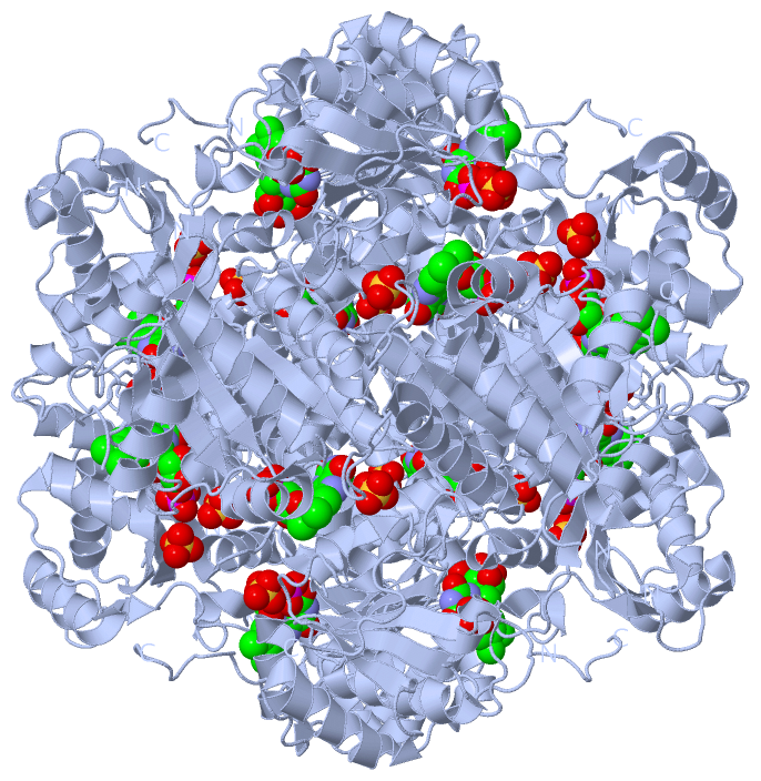
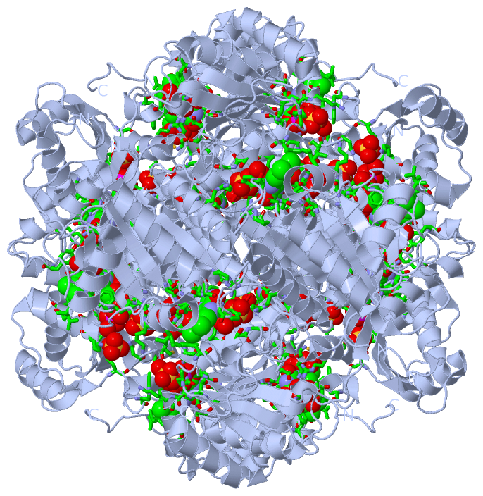
Ligands, Modified Residues, Ions (2, 3)| Asymmetric Unit (2, 3) Biological Unit 1 (2, 36) |
Sites (3, 3)
Asymmetric Unit (3, 3)
|
SS Bonds (0, 0)| (no "SS Bond" information available for 3ZQU) |
Cis Peptide Bonds (2, 2)
Asymmetric Unit
|
||||||||||||
SAPs(SNPs)/Variants (0, 0)| (no "SAP(SNP)/Variant" information available for 3ZQU) |
PROSITE Motifs (0, 0)| (no "PROSITE Motif" information available for 3ZQU) |
Exons (0, 0)| (no "Exon" information available for 3ZQU) |
Sequences/Alignments
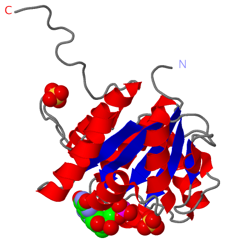
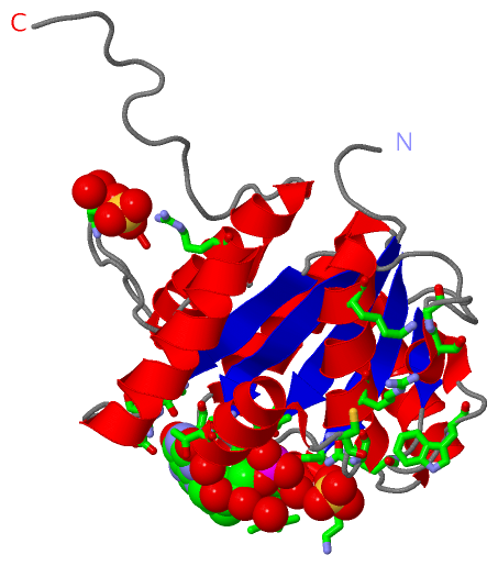
Asymmetric UnitChain A from PDB Type:PROTEIN Length:207 aligned with UBIX_PSEAE | Q9HX08 from UniProtKB/Swiss-Prot Length:209 Alignment length:207 10 20 30 40 50 60 70 80 90 100 110 120 130 140 150 160 170 180 190 200 UBIX_PSEAE 1 MSGPERITLAMTGASGAQYGLRLLDCLVQEEREVHFLISKAAQLVMATETDVALPAKPQAMQAFLTEYCGAAAGQIRVFGQNDWMAPPASGSSAPNAMVICPCSTGTLSAVATGACNNLIERAADVALKERRPLVLVPREAPFSSIHLENMLKLSNLGAVILPAAPGFYHQPQSVEDLVDFVVARILNTLGIPQDMLPRWGEQHLVS 207 SCOP domains d3zqua_ A: automated matches SCOP domains CATH domains --------------------------------------------------------------------------------------------------------------------------------------------------------------------------------------------------------------- CATH domains Pfam domains --------------------------------------------------------------------------------------------------------------------------------------------------------------------------------------------------------------- Pfam domains SAPs(SNPs) --------------------------------------------------------------------------------------------------------------------------------------------------------------------------------------------------------------- SAPs(SNPs) PROSITE --------------------------------------------------------------------------------------------------------------------------------------------------------------------------------------------------------------- PROSITE Transcript --------------------------------------------------------------------------------------------------------------------------------------------------------------------------------------------------------------- Transcript 3zqu A 1 MSGPERITLAMTGASGAQYGLRLLDCLVQEEREVHFLISKAAQLVMATETDVALPAKPQAMQAFLTEYCGAAAGQIRVFGQNDWMAPPASGSSAPNAMVICPCSTGTLSAVATGACNNLIERAADVALKERRPLVLVPREAPFSSIHLENMLKLSNLGAVILPAAPGFYHQPQSVEDLVDFVVARILNTLGIPQDMLPRWGEQHLVS 207 10 20 30 40 50 60 70 80 90 100 110 120 130 140 150 160 170 180 190 200
|
||||||||||||||||||||
SCOP Domains (1, 1)
Asymmetric Unit
 |
CATH Domains (0, 0)| (no "CATH Domain" information available for 3ZQU) |
Pfam Domains (0, 0)| (no "Pfam Domain" information available for 3ZQU) |
Gene Ontology (4, 4)|
Asymmetric Unit(hide GO term definitions) Chain A (UBIX_PSEAE | Q9HX08)
|
||||||||||||||||||||||||||||||||||||
Interactive Views
|
|||||||||||||||||||||||||||||||||||||||||||||||||||||||||||||||||||||||||||||||||||||||||||||||||||||||||||||||||||||||||||||||||||||||||||||||||||||||||||||||||||||
Still Images
|
||||||||||||||||
Databases
|
||||||||||||||||||||||||||||||||||||||||||||||||||||||||||||||||||||||||||||||||||||||||||||||||||||||||||||||||||||||||||||||||||||||||||||||||||||||||||||||||
Analysis Tools
|
|||||||||||||||||||||||||||||||||||||||||||||||||||||||||||||
Entries Sharing at Least One Protein Chain (UniProt ID)
Related Entries Specified in the PDB File
|
|