Show PDB file:   
         Plain Text   HTML   (compressed file size)
QuickSearch:   
by PDB,NDB,UniProt,PROSITE Code or Search Term(s)  
(-)Asymmetric Unit
(-)Asym. Unit - sites
(-)Biological Unit 1
(-)Biol. Unit 1 - sites
(-)Biological Unit 2
collapse expand < >
Image Asymmetric Unit
Asymmetric Unit  (Jmol Viewer)
Image Asym. Unit - sites
Asym. Unit - sites  (Jmol Viewer)
Image Biological Unit 1
Biological Unit 1  (Jmol Viewer)
Image Biol. Unit 1 - sites
Biol. Unit 1 - sites  (Jmol Viewer)
Image Biological Unit 2
Biological Unit 2  (Jmol Viewer)

(-) Description

Title :  CRYSTAL STRUCTURE OF DNA URIDINE ENDONUCLEASE MTH212 MUTANT W205S
 
Authors :  N. Tabata, T. Shida, R. Arai
Date :  06 Dec 12  (Deposition) - 11 Dec 13  (Release) - 11 Dec 13  (Revision)
Method :  X-RAY DIFFRACTION
Resolution :  1.90
Chains :  Asym. Unit :  A,D
Biol. Unit 1:  A  (1x)
Biol. Unit 2:  D  (1x)
Keywords :  Alpha/Beta-Sandwich, Hydrolase, Dna Binding (Keyword Search: [Gene Ontology, PubMed, Web (Google))
 
Reference :  N. Tabata, T. Shida, R. Arai
Crystal Structure Of Dna Uridine Endonuclease Mth212
To Be Published
PubMed: search

(-) Compounds

Molecule 1 - EXODEOXYRIBONUCLEASE
    ChainsA, D
    EC Number3.1.11.2
    EngineeredYES
    Expression SystemESCHERICHIA COLI
    Expression System PlasmidPET15B
    Expression System StrainBL21(DE3)
    Expression System Taxid562
    Expression System Vector TypePLASMID
    GeneMTH212, MTH_212
    MutationYES
    Organism ScientificMETHANOTHERMOBACTER THERMAUTOTROPHICUS
    Organism Taxid187420
    StrainATCC 29096 / DSM 1053 / JCM 10044 / NBRC 100330 / DELTA H

 Structural Features

(-) Chains, Units

  12
Asymmetric Unit AD
Biological Unit 1 (1x)A 
Biological Unit 2 (1x) D

Summary Information (see also Sequences/Alignments below)

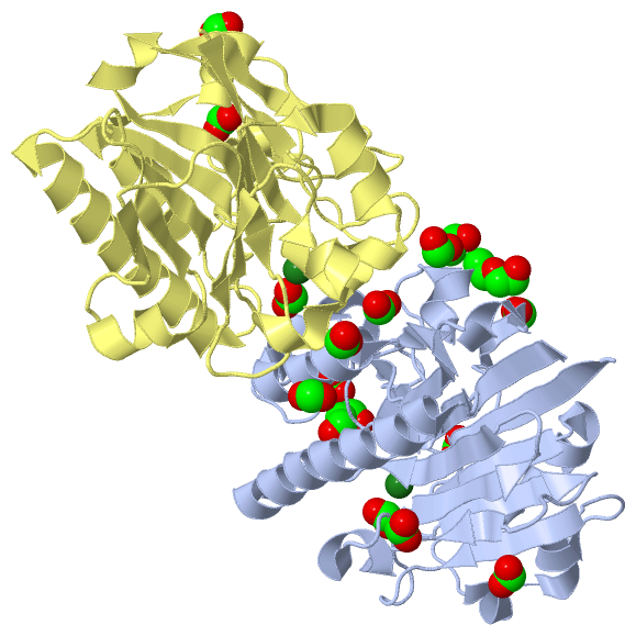
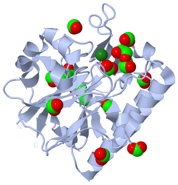
(-) Ligands, Modified Residues, Ions  (3, 17)

Asymmetric Unit (3, 17)
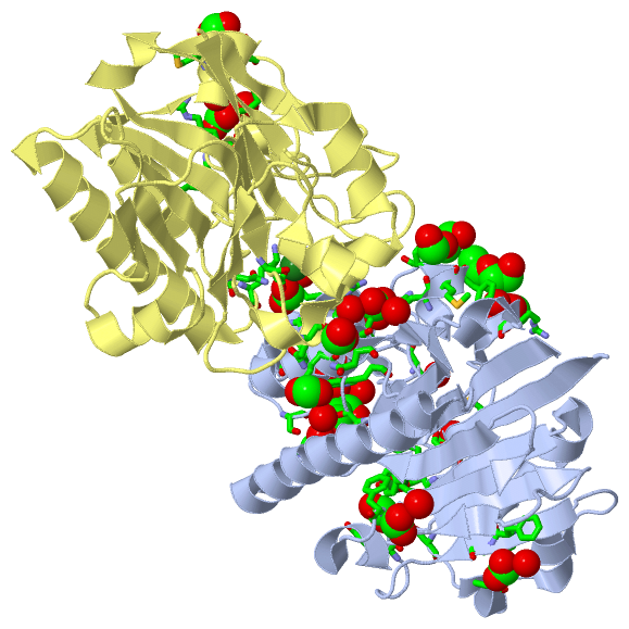
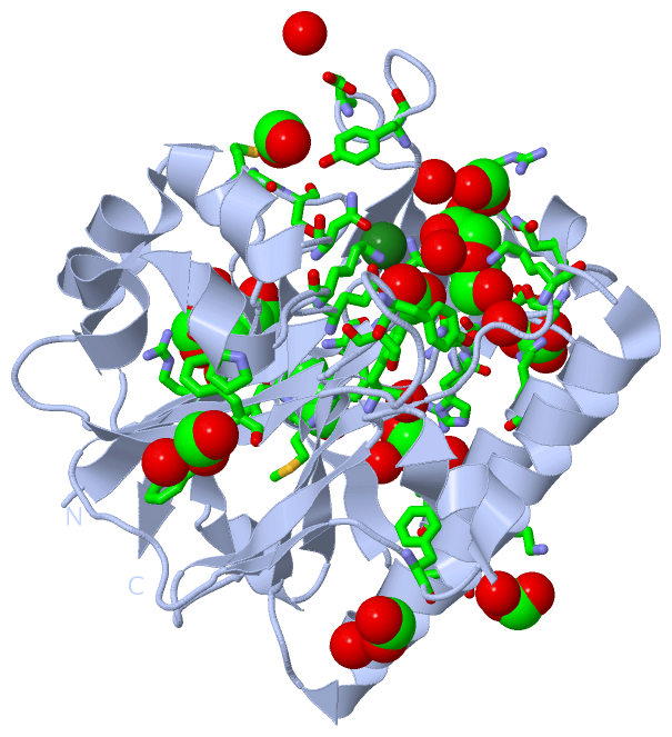
No.NameCountTypeFull Name
1FMT12Ligand/IonFORMIC ACID
2MG2Ligand/IonMAGNESIUM ION
3PEG3Ligand/IonDI(HYDROXYETHYL)ETHER
Biological Unit 1 (2, 12)
No.NameCountTypeFull Name
1FMT9Ligand/IonFORMIC ACID
2MG-1Ligand/IonMAGNESIUM ION
3PEG3Ligand/IonDI(HYDROXYETHYL)ETHER
Biological Unit 2 (1, 3)
No.NameCountTypeFull Name
1FMT3Ligand/IonFORMIC ACID
2MG-1Ligand/IonMAGNESIUM ION
3PEG-1Ligand/IonDI(HYDROXYETHYL)ETHER

(-) Sites  (15, 15)

Asymmetric Unit (15, 15)
No.NameEvidenceResiduesDescription
01AC1SOFTWAREASN A:12 , GLU A:38 , HOH A:411 , HOH A:468 , HOH A:509 , HOH A:590BINDING SITE FOR RESIDUE MG A 301
02AC2SOFTWAREPHE A:86 , HOH A:594BINDING SITE FOR RESIDUE FMT A 302
03AC3SOFTWARELYS A:183 , ASN A:187 , HOH A:495 , GLN D:45BINDING SITE FOR RESIDUE FMT A 303
04AC4SOFTWAREPRO A:43 , PHE A:59 , PHE A:60 , GLU A:214 , HOH A:431 , HOH A:558BINDING SITE FOR RESIDUE FMT A 304
05AC5SOFTWAREGLU A:158 , ARG A:178 , HOH A:613 , ASN D:114 , LYS D:116BINDING SITE FOR RESIDUE FMT A 305
06AC6SOFTWAREARG A:236 , HOH A:447 , HOH A:614BINDING SITE FOR RESIDUE FMT A 306
07AC7SOFTWARELYS A:116 , ASN A:153 , SER A:171 , TRP A:219 , HOH A:413 , HOH A:579BINDING SITE FOR RESIDUE PEG A 308
08AC8SOFTWARETYR A:208 , MET A:244 , GLY A:245 , SER A:246BINDING SITE FOR RESIDUE FMT A 309
09AC9SOFTWAREARG A:194 , MET A:195 , PHE A:196 , SER A:198BINDING SITE FOR RESIDUE PEG A 310
10BC1SOFTWARELYS A:40 , TYR A:68 , SER A:69 , TYR A:111BINDING SITE FOR RESIDUE FMT A 311
11BC2SOFTWAREARG A:163 , ASN A:167 , TRP A:219 , HOH A:435 , HOH A:479BINDING SITE FOR RESIDUE FMT A 312
12BC3SOFTWAREHIS A:156 , ARG A:157 , ILE A:185 , HOH A:430 , HOH A:456 , HOH A:581BINDING SITE FOR RESIDUE FMT A 313
13BC4SOFTWAREASN D:12 , GLU D:38 , HOH D:407 , HOH D:425 , HOH D:430 , HOH D:449BINDING SITE FOR RESIDUE MG D 301
14BC5SOFTWAREARG D:157 , GLU D:158 , ILE D:159 , ARG D:178 , HOH D:525 , HOH D:535BINDING SITE FOR RESIDUE FMT D 302
15BC6SOFTWAREMET D:195 , SER D:198BINDING SITE FOR RESIDUE FMT D 303

(-) SS Bonds  (0, 0)

(no "SS Bond" information available for 3W2Y)

(-) Cis Peptide Bonds  (0, 0)

(no "Cis Peptide Bond" information available for 3W2Y)

 Sequence-Structure Mapping

(-) SAPs(SNPs)/Variants  (0, 0)

(no "SAP(SNP)/Variant" information available for 3W2Y)

(-) PROSITE Motifs  (0, 0)

(no "PROSITE Motif" information available for 3W2Y)

(-) Exons   (0, 0)

(no "Exon" information available for 3W2Y)

(-) Sequences/Alignments

Asymmetric Unit
   Reformat: Number of residues per line =  ('0' or empty: single-line sequence representation)
  Number of residues per labelling interval =   
  UniProt sequence: complete  aligned part    
   Show mapping: SCOP domains CATH domains Pfam domains Secondary structure (by author)
SAPs(SNPs) PROSITE motifs Exons
(details for a mapped element are shown in a popup box when the mouse pointer rests over it)
Chain A from PDB  Type:PROTEIN  Length:256
 aligned with O26314_METTH | O26314 from UniProtKB/TrEMBL  Length:257

    Alignment length:256
                                    11        21        31        41        51        61        71        81        91       101       111       121       131       141       151       161       171       181       191       201       211       221       231       241       251      
         O26314_METTH     2 TVLKIISWNVNGLRAVHRKGFLKWFMEEKPDILCLQEIKAAPEQLPRKLRHVEGYRSFFTPAERKGYSGVAMYTKVPPSSLREGFGVERFDTEGRIQIADFDDFLLYNIYFPNGKMSEERLKYKLEFYDAFLEDVNRERDSGRNVIICGDFNTAHREIDLARPKENSNVSGFLPVERAWIDKFIENGYVDTFRMFNSDPGQYTWWSYRTRARERNVGWRLDYFFVNEEFKGKVKRSWILSDVMGSDHCPIGLEIEL 257
               SCOP domains d3w2ya_ A: automated matches                                                                                                                                                                                                                                     SCOP domains
               CATH domains ---------------------------------------------------------------------------------------------------------------------------------------------------------------------------------------------------------------------------------------------------------------- CATH domains
               Pfam domains ---------------------------------------------------------------------------------------------------------------------------------------------------------------------------------------------------------------------------------------------------------------- Pfam domains
         Sec.struct. author .eeeeeeee..hhhhhhh.hhhhhhhhhh..eeeee....hhhhhhhhhhh....eeeee.........eeeee......ee....hhhhhh...eeeee....eeeeee......hhhhhhhhhhhhhhhhhhhhhhhhh...eeeeee.....hhhhh.hhhhhh.....hhhhhhhhhhhhhh.eeehhhhhh.......ee...........ee..eeeeee.hhhh.eeeeee...........eeeeee. Sec.struct. author
                 SAPs(SNPs) ---------------------------------------------------------------------------------------------------------------------------------------------------------------------------------------------------------------------------------------------------------------- SAPs(SNPs)
                    PROSITE ---------------------------------------------------------------------------------------------------------------------------------------------------------------------------------------------------------------------------------------------------------------- PROSITE
                 Transcript ---------------------------------------------------------------------------------------------------------------------------------------------------------------------------------------------------------------------------------------------------------------- Transcript
                 3w2y A   2 TVLKIISWNVNGLRAVHRKGFLKWFMEEKPDILCLQEIKAAPEQLPRKLRHVEGYRSFFTPAERKGYSGVAMYTKVPPSSLREGFGVERFDTEGRIQIADFDDFLLYNIYFPNGKMSEERLKYKLEFYDAFLEDVNRERDSGRNVIICGDFNTAHREIDLARPKENSNVSGFLPVERAWIDKFIENGYVDTFRMFNSDPGQYTSWSYRTRARERNVGWRLDYFFVNEEFKGKVKRSWILSDVMGSDHCPIGLEIEL 257
                                    11        21        31        41        51        61        71        81        91       101       111       121       131       141       151       161       171       181       191       201       211       221       231       241       251      

Chain D from PDB  Type:PROTEIN  Length:256
 aligned with O26314_METTH | O26314 from UniProtKB/TrEMBL  Length:257

    Alignment length:256
                                    11        21        31        41        51        61        71        81        91       101       111       121       131       141       151       161       171       181       191       201       211       221       231       241       251      
         O26314_METTH     2 TVLKIISWNVNGLRAVHRKGFLKWFMEEKPDILCLQEIKAAPEQLPRKLRHVEGYRSFFTPAERKGYSGVAMYTKVPPSSLREGFGVERFDTEGRIQIADFDDFLLYNIYFPNGKMSEERLKYKLEFYDAFLEDVNRERDSGRNVIICGDFNTAHREIDLARPKENSNVSGFLPVERAWIDKFIENGYVDTFRMFNSDPGQYTWWSYRTRARERNVGWRLDYFFVNEEFKGKVKRSWILSDVMGSDHCPIGLEIEL 257
               SCOP domains d3w2yd_ D: automated matches                                                                                                                                                                                                                                     SCOP domains
               CATH domains ---------------------------------------------------------------------------------------------------------------------------------------------------------------------------------------------------------------------------------------------------------------- CATH domains
               Pfam domains ---------------------------------------------------------------------------------------------------------------------------------------------------------------------------------------------------------------------------------------------------------------- Pfam domains
         Sec.struct. author .eeeeeeee..hhhhhhh.hhhhhhhhhh..eeeee....hhhhhhhhhhh....eeeee.........eeeee.....eee....hhhhhh...eeeee....eeeeee......hhhhhhhhhhhhhhhhhhhhhhhhh...eeeeee.....hhhhh............hhhhhhhhhhhhhh.eeehhhhhh........................eeeeee.hhhh.eeeeee...........eeeeee. Sec.struct. author
                 SAPs(SNPs) ---------------------------------------------------------------------------------------------------------------------------------------------------------------------------------------------------------------------------------------------------------------- SAPs(SNPs)
                    PROSITE ---------------------------------------------------------------------------------------------------------------------------------------------------------------------------------------------------------------------------------------------------------------- PROSITE
                 Transcript ---------------------------------------------------------------------------------------------------------------------------------------------------------------------------------------------------------------------------------------------------------------- Transcript
                 3w2y D   2 TVLKIISWNVNGLRAVHRKGFLKWFMEEKPDILCLQEIKAAPEQLPRKLRHVEGYRSFFTPAERKGYSGVAMYTKVPPSSLREGFGVERFDTEGRIQIADFDDFLLYNIYFPNGKMSEERLKYKLEFYDAFLEDVNRERDSGRNVIICGDFNTAHREIDLARPKENSNVSGFLPVERAWIDKFIENGYVDTFRMFNSDPGQYTSWSYRTRARERNVGWRLDYFFVNEEFKGKVKRSWILSDVMGSDHCPIGLEIEL 257
                                    11        21        31        41        51        61        71        81        91       101       111       121       131       141       151       161       171       181       191       201       211       221       231       241       251      

   Legend:   → Mismatch (orange background)
  - → Gap (green background, '-', border residues have a numbering label)
    → Modified Residue (blue background, lower-case, 'x' indicates undefined single-letter code, labelled with number + name)
  x → Chemical Group (purple background, 'x', labelled with number + name, e.g. ACE or NH2)
  extra numbering lines below/above indicate numbering irregularities and modified residue names etc., number ends below/above '|'

 Classification and Annotation

(-) SCOP Domains  (1, 2)

Asymmetric Unit

(-) CATH Domains  (0, 0)

(no "CATH Domain" information available for 3W2Y)

(-) Pfam Domains  (0, 0)

(no "Pfam Domain" information available for 3W2Y)

(-) Gene Ontology  (7, 7)

Asymmetric Unit(hide GO term definitions)
Chain A,D   (O26314_METTH | O26314)
molecular function
    GO:0003677    DNA binding    Any molecular function by which a gene product interacts selectively and non-covalently with DNA (deoxyribonucleic acid).
    GO:0004519    endonuclease activity    Catalysis of the hydrolysis of ester linkages within nucleic acids by creating internal breaks.
    GO:0046872    metal ion binding    Interacting selectively and non-covalently with any metal ion.
    GO:0004518    nuclease activity    Catalysis of the hydrolysis of ester linkages within nucleic acids.
biological process
    GO:0006281    DNA repair    The process of restoring DNA after damage. Genomes are subject to damage by chemical and physical agents in the environment (e.g. UV and ionizing radiations, chemical mutagens, fungal and bacterial toxins, etc.) and by free radicals or alkylating agents endogenously generated in metabolism. DNA is also damaged because of errors during its replication. A variety of different DNA repair pathways have been reported that include direct reversal, base excision repair, nucleotide excision repair, photoreactivation, bypass, double-strand break repair pathway, and mismatch repair pathway.
    GO:0090305    nucleic acid phosphodiester bond hydrolysis    The nucleic acid metabolic process in which the phosphodiester bonds between nucleotides are cleaved by hydrolysis.
cellular component
    GO:0005622    intracellular    The living contents of a cell; the matter contained within (but not including) the plasma membrane, usually taken to exclude large vacuoles and masses of secretory or ingested material. In eukaryotes it includes the nucleus and cytoplasm.

 Visualization

(-) Interactive Views

Asymmetric Unit
  Complete Structure
    Jena3D(integrated viewing of ligand, site, SAP, PROSITE, SCOP information)
    WebMol | AstexViewer[tm]@PDBe
(Java Applets, require no local installation except for Java; loading may be slow)
    STRAP
(Java WebStart application, automatic local installation, requires Java; full application with system access!)
    RasMol
(require local installation)
    Molscript (VRML)
(requires installation of a VRML viewer; select preferred view via VRML and generate a mono or stereo PDF format file)
 
  Ligands, Modified Residues, Ions
    FMT  [ RasMol | Jena3D ]  +environment [ RasMol | Jena3D ]
    MG  [ RasMol | Jena3D ]  +environment [ RasMol | Jena3D ]
    PEG  [ RasMol | Jena3D ]  +environment [ RasMol | Jena3D ]
 
  Sites
    AC1  [ RasMol ]  +environment [ RasMol ]
    AC2  [ RasMol ]  +environment [ RasMol ]
    AC3  [ RasMol ]  +environment [ RasMol ]
    AC4  [ RasMol ]  +environment [ RasMol ]
    AC5  [ RasMol ]  +environment [ RasMol ]
    AC6  [ RasMol ]  +environment [ RasMol ]
    AC7  [ RasMol ]  +environment [ RasMol ]
    AC8  [ RasMol ]  +environment [ RasMol ]
    AC9  [ RasMol ]  +environment [ RasMol ]
    BC1  [ RasMol ]  +environment [ RasMol ]
    BC2  [ RasMol ]  +environment [ RasMol ]
    BC3  [ RasMol ]  +environment [ RasMol ]
    BC4  [ RasMol ]  +environment [ RasMol ]
    BC5  [ RasMol ]  +environment [ RasMol ]
    BC6  [ RasMol ]  +environment [ RasMol ]
 
  Cis Peptide Bonds
(no "Cis Peptide Bonds" information available for 3w2y)
 
Biological Units
  Complete Structure
    Biological Unit 1  [ Jena3D ]
    Biological Unit 2  [ Jena3D ]

(-) Still Images

Jmol
  protein: cartoon or spacefill or dots and stick; nucleic acid: cartoon and stick; ligands: spacefill; active site: stick
Molscript
  protein, nucleic acid: cartoon; ligands: spacefill; active site: ball and stick

 Databases and Analysis Tools

(-) Databases

Access by PDB/NDB ID
  3w2y
    Family and Domain InformationProDom | SYSTERS
    General Structural InformationGlycoscienceDB | MMDB | NDB | OCA | PDB | PDBe | PDBj | PDBsum | PDBWiki | PQS | PROTEOPEDIA
    Orientation in MembranesOPM
    Protein SurfaceSURFACE
    Secondary StructureDSSP (structure derived) | HSSP (homology derived)
    Structural GenomicsGeneCensus
    Structural NeighboursCE | VAST
    Structure ClassificationCATH | Dali | SCOP
    Validation and Original DataBMRB Data View | BMRB Restraints Grid | EDS | PROCHECK | RECOORD | WHAT_CHECK
 
Access by UniProt ID/Accession number
  O26314_METTH | O26314
    Comparative Protein Structure ModelsModBase
    Genomic InformationEnsembl
    Protein-protein InteractionDIP
    Sequence, Family and Domain InformationInterPro | Pfam | SMART | UniProtKB/TrEMBL
 
Access by Enzyme Classificator   (EC Number)
  3.1.11.2
    General Enzyme InformationBRENDA | EC-PDB | Enzyme | IntEnz
    PathwayKEGG | MetaCyc
 
Access by Disease Identifier   (MIM ID)
  (no 'MIM ID' available)
    Disease InformationOMIM
 
Access by GenAge ID
  (no 'GenAge ID' available)
    Age Related InformationGenAge

(-) Analysis Tools

Access by PDB/NDB ID
    Domain InformationXDom
    Interatomic Contacts of Structural UnitsCSU
    Ligand-protein ContactsLPC
    Protein CavitiescastP
    Sequence and Secondary StructurePDBCartoon
    Structure AlignmentSTRAP(Java WebStart application, automatic local installation, requires Java; full application with system access!)
    Structure and Sequence BrowserSTING
 
Access by UniProt ID/Accession number
  O26314_METTH | O26314
    Protein Disorder PredictionDisEMBL | FoldIndex | GLOBPLOT (for more information see DisProt)

 Related Entries

(-) Entries Sharing at Least One Protein Chain (UniProt ID)

UniProtKB/TrEMBL
        O26314_METTH | O263143fzi 3g00 3g0a 3g0r 3g1k 3g2c 3g2d 3g38 3g3c 3g3y 3g4t 3g8v 3g91 3ga6 3w2x

(-) Related Entries Specified in the PDB File

3w2x THE WILD TYPE ENZYME