Show PDB file:   
         Plain Text   HTML   (compressed file size)
QuickSearch:   
by PDB,NDB,UniProt,PROSITE Code or Search Term(s)  
(-)Asym./Biol. Unit
(-)Asym./Biol. Unit - sites
collapse expand < >
Image Asym./Biol. Unit
Asym./Biol. Unit  (Jmol Viewer)
Image Asym./Biol. Unit - sites
Asym./Biol. Unit - sites  (Jmol Viewer)

(-) Description

Title :  CRYSTAL STRUCTURE OF RAT VITAMIN D RECEPTOR BOUND TO A PARTIAL AGONIST 26-ADAMANTYL-23-YNE-19-NORVITAMMIN D ADTK3
 
Authors :  M. Nakabayashi, T. Kudo, H. Tokiwa, M. Makishima, S. Yamada, T. Ikura, N
Date :  26 May 12  (Deposition) - 12 Jun 13  (Release) - 14 May 14  (Revision)
Method :  X-RAY DIFFRACTION
Resolution :  1.50
Chains :  Asym./Biol. Unit :  A,B
Keywords :  Transcription, Hormone Receptor (Keyword Search: [Gene Ontology, PubMed, Web (Google))
 
Reference :  T. Kudo, M. Ishizawa, K. Maekawa, M. Nakabayashi, Y. Watarai, H. Uchida, H. Tokiwa, T. Ikura, N. Ito, M. Makishima, S. Yamada
Combination Of Triple Bond And Adamantane Ring On The Vitamin D Side Chain Produced Partial Agonists For Vitamin Receptor.
J. Med. Chem. 2014
PubMed-ID: 24773565  |  Reference-DOI: 10.1021/JM401989C

(-) Compounds

Molecule 1 - VITAMIN D3 RECEPTOR
    ChainsA
    EngineeredYES
    Expression SystemESCHERICHIA COLI
    Expression System PlasmidPET14B
    Expression System StrainC41
    Expression System Taxid562
    Expression System Vector TypePLASMID
    FragmentUNP RESIDUES 116-423
    GeneNR1I1, VDR
    Organism CommonRATS
    Organism ScientificRATTUS NORVEGICUS
    Organism Taxid10116
    SynonymVDR, 1,25-DIHYDROXYVITAMIN D3 RECEPTOR, NUCLEAR RECEPTOR SUBFAMILY 1 GROUP I MEMBER 1
 
Molecule 2 - COACTIVATOR PEPTIDE DRIP
    ChainsB
    EngineeredYES
    Other DetailsPEPTIDE SYNTHESIS
    SyntheticYES

 Structural Features

(-) Chains, Units

  12
Asymmetric/Biological Unit AB

Summary Information (see also Sequences/Alignments below)

(-) Ligands, Modified Residues, Ions  (2, 2)

Asymmetric/Biological Unit (2, 2)
No.NameCountTypeFull Name
1EDO1Ligand/Ion1,2-ETHANEDIOL
2TK31Ligand/Ion(1R,3R,7E,17BETA)-17-{(2R,6R)-6-HYDROXY-7-[(3S,5S,7S)-TRICYCLO[3.3.1.1~3,7~]DEC-1-YL]HEPT-4-YN-2-YL}-2-METHYLIDENE-9,10-SECOESTRA-5,7-DIENE-1,3-DIOL

(-) Sites  (2, 2)

Asymmetric Unit (2, 2)
No.NameEvidenceResiduesDescription
1AC1SOFTWARETYR A:143 , LEU A:223 , LEU A:226 , ALA A:227 , VAL A:230 , SER A:233 , ARG A:270 , SER A:271 , SER A:274 , TRP A:282 , CYS A:284 , HIS A:301 , HIS A:393 , TYR A:397 , LEU A:400 , LEU A:410BINDING SITE FOR RESIDUE TK3 A 500
2AC2SOFTWAREASP A:137 , LEU A:347 , ILE A:351 , ARG A:354 , HOH A:614 , HOH A:623 , HOH A:631BINDING SITE FOR RESIDUE EDO A 501

(-) SS Bonds  (0, 0)

(no "SS Bond" information available for 3VTC)

(-) Cis Peptide Bonds  (1, 1)

Asymmetric/Biological Unit
No.Residues
1Pro A:369 -Pro A:370

 Sequence-Structure Mapping

(-) SAPs(SNPs)/Variants  (0, 0)

(no "SAP(SNP)/Variant" information available for 3VTC)

(-) PROSITE Motifs  (0, 0)

(no "PROSITE Motif" information available for 3VTC)

(-) Exons   (0, 0)

(no "Exon" information available for 3VTC)

(-) Sequences/Alignments

Asymmetric/Biological Unit
   Reformat: Number of residues per line =  ('0' or empty: single-line sequence representation)
  Number of residues per labelling interval =   
  UniProt sequence: complete  aligned part    
   Show mapping: SCOP domains CATH domains Pfam domains Secondary structure (by author)
SAPs(SNPs) PROSITE motifs Exons
(details for a mapped element are shown in a popup box when the mouse pointer rests over it)
Chain A from PDB  Type:PROTEIN  Length:240
 aligned with VDR_RAT | P13053 from UniProtKB/Swiss-Prot  Length:423

    Alignment length:298
                                   132       142       152       162       172       182       192       202       212       222       232       242       252       262       272       282       292       302       312       322       332       342       352       362       372       382       392       402       412        
              VDR_RAT   123 KLSEEQQHIIAILLDAHHKTYDPTYADFRDFRPPVRMDGSTGSYSPRPTLSFSGNSSSSSSDLYTTSLDMMEPSGFSNLDLNGEDSDDPSVTLDLSPLSMLPHLADLVSYSIQKVIGFAKMIPGFRDLTSDDQIVLLKSSAIEVIMLRSNQSFTMDDMSWDCGSQDYKYDVTDVSKAGHTLELIEPLIKFQVGLKKLNLHEEEHVLLMAICIVSPDRPGVQDAKLVEAIQDRLSNTLQTYIRCRHPPPGSHQLYAKMIQKLADLRSLNEEHSKQYRSLSFQPENSMKLTPLVLEVFGN 420
               SCOP domains ---------------------------------------------------------------------------------------------------------------------------------------------------------------------------------------------------------------------------------------------------------------------------------------------------------- SCOP domains
               CATH domains ---------------------------------------------------------------------------------------------------------------------------------------------------------------------------------------------------------------------------------------------------------------------------------------------------------- CATH domains
               Pfam domains ---------------------------------------------------------------------------------------------------------------------------------------------------------------------------------------------------------------------------------------------------------------------------------------------------------- Pfam domains
         Sec.struct. author ..hhhhhhhhhhhhhhhhhhh...hhhhhhh......----------------------------------------------------------....hhhhhhhhhhhhhhhhhhhhh...hhhhhhhhhhhhhhhhhhhhhhhhhh...ee....eee..hhh.eehhhhhhh...hhhhhhhhhhhhhhhhh...hhhhhhhhhhhhhh........hhhhhhhhhhhhhhhhhhhhhhhh......hhhhhhhhhhhhhhhhhhhhhhhhhhhhhhhhhhh..hhhhhhhhh. Sec.struct. author
                 SAPs(SNPs) ---------------------------------------------------------------------------------------------------------------------------------------------------------------------------------------------------------------------------------------------------------------------------------------------------------- SAPs(SNPs)
                    PROSITE ---------------------------------------------------------------------------------------------------------------------------------------------------------------------------------------------------------------------------------------------------------------------------------------------------------- PROSITE
                 Transcript ---------------------------------------------------------------------------------------------------------------------------------------------------------------------------------------------------------------------------------------------------------------------------------------------------------- Transcript
                 3vtc A 123 KLSEEQQHIIAILLDAHHKTYDPTYADFRDFRPPVRM----------------------------------------------------------SPLSMLPHLADLVSYSIQKVIGFAKMIPGFRDLTSDDQIVLLKSSAIEVIMLRSNQSFTMDDMSWDCGSQDYKYDVTDVSKAGHTLELIEPLIKFQVGLKKLNLHEEEHVLLMAICIVSPDRPGVQDAKLVEAIQDRLSNTLQTYIRCRHPPPGSHQLYAKMIQKLADLRSLNEEHSKQYRSLSFQPENSMKLTPLVLEVFGN 420
                                   132       142       152      |  -         -         -         -         -         -     | 222       232       242       252       262       272       282       292       302       312       322       332       342       352       362       372       382       392       402       412        
                                                              159                                                        218                                                                                                                                                                                                          

Chain B from PDB  Type:PROTEIN  Length:11
                                           
               SCOP domains ----------- SCOP domains
               CATH domains ----------- CATH domains
               Pfam domains ----------- Pfam domains
         Sec.struct. author ..hhhhhhhhh Sec.struct. author
                 SAPs(SNPs) ----------- SAPs(SNPs)
                    PROSITE ----------- PROSITE
                 Transcript ----------- Transcript
                 3vtc B 625 KNHPMLMNLLK 635
                                   634 

   Legend:   → Mismatch (orange background)
  - → Gap (green background, '-', border residues have a numbering label)
    → Modified Residue (blue background, lower-case, 'x' indicates undefined single-letter code, labelled with number + name)
  x → Chemical Group (purple background, 'x', labelled with number + name, e.g. ACE or NH2)
  extra numbering lines below/above indicate numbering irregularities and modified residue names etc., number ends below/above '|'

 Classification and Annotation

(-) SCOP Domains  (0, 0)

(no "SCOP Domain" information available for 3VTC)

(-) CATH Domains  (0, 0)

(no "CATH Domain" information available for 3VTC)

(-) Pfam Domains  (0, 0)

(no "Pfam Domain" information available for 3VTC)

(-) Gene Ontology  (33, 33)

Asymmetric/Biological Unit(hide GO term definitions)
Chain A   (VDR_RAT | P13053)
molecular function
    GO:0003677    DNA binding    Any molecular function by which a gene product interacts selectively and non-covalently with DNA (deoxyribonucleic acid).
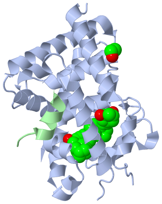
    GO:1902098    calcitriol binding    Interacting selectively and non-covalently with calcitriol. Calcitriol (1,25-dihydroxycholecalciferol) is the hormonally active form of vitamin D3.
    GO:0008434    calcitriol receptor activity    Combining with calcitriol, the hormonally active form of vitamin D3, and transmitting the signal to the transcriptional machinery by interacting selectively and non-covalently with a specific DNA sequence in order to modulate transcription by RNA polymerase II.
    GO:0046872    metal ion binding    Interacting selectively and non-covalently with any metal ion.
    GO:0005515    protein binding    Interacting selectively and non-covalently with any protein or protein complex (a complex of two or more proteins that may include other nonprotein molecules).
    GO:0043565    sequence-specific DNA binding    Interacting selectively and non-covalently with DNA of a specific nucleotide composition, e.g. GC-rich DNA binding, or with a specific sequence motif or type of DNA e.g. promotor binding or rDNA binding.
    GO:0003707    steroid hormone receptor activity    Combining with a steroid hormone and transmitting the signal within the cell to initiate a change in cell activity or function.
    GO:0003700    transcription factor activity, sequence-specific DNA binding    Interacting selectively and non-covalently with a specific DNA sequence in order to modulate transcription. The transcription factor may or may not also interact selectively with a protein or macromolecular complex.
    GO:0005499    vitamin D binding    Interacting selectively and non-covalently with vitamin D, any of a group of related, fat-soluble compounds that are derived from delta-5,7 steroids and play a central role in calcium metabolism. Specific forms of vitamin D include calciferol (ergocalciferol; vitamin D2) and cholecalciferol (calciol; vitamin D3).
    GO:0008270    zinc ion binding    Interacting selectively and non-covalently with zinc (Zn) ions.
biological process
    GO:0007568    aging    A developmental process that is a deterioration and loss of function over time. Aging includes loss of functions such as resistance to disease, homeostasis, and fertility, as well as wear and tear. Aging includes cellular senescence, but is more inclusive. May precede death and may succeed developmental maturation (GO:0021700).
    GO:0097190    apoptotic signaling pathway    A series of molecular signals which triggers the apoptotic death of a cell. The pathway starts with reception of a signal, and ends when the execution phase of apoptosis is triggered.
    GO:0071305    cellular response to vitamin D    Any process that results in a change in state or activity of a cell (in terms of movement, secretion, enzyme production, gene expression, etc.) as a result of a vitamin D stimulus.
    GO:0007507    heart development    The process whose specific outcome is the progression of the heart over time, from its formation to the mature structure. The heart is a hollow, muscular organ, which, by contracting rhythmically, keeps up the circulation of the blood.
    GO:0051924    regulation of calcium ion transport    Any process that modulates the frequency, rate or extent of the directed movement of calcium ions into, out of or within a cell, or between cells, by means of some agent such as a transporter or pore.
    GO:0006357    regulation of transcription from RNA polymerase II promoter    Any process that modulates the frequency, rate or extent of transcription from an RNA polymerase II promoter.
    GO:0006355    regulation of transcription, DNA-templated    Any process that modulates the frequency, rate or extent of cellular DNA-templated transcription.
    GO:0051592    response to calcium ion    Any process that results in a change in state or activity of a cell or an organism (in terms of movement, secretion, enzyme production, gene expression, etc.) as a result of a calcium ion stimulus.
    GO:0032355    response to estradiol    Any process that results in a change in state or activity of a cell or an organism (in terms of movement, secretion, enzyme production, gene expression, etc.) as a result of stimulus by estradiol, a C18 steroid hormone hydroxylated at C3 and C17 that acts as a potent estrogen.
    GO:0043401    steroid hormone mediated signaling pathway    A series of molecular signals mediated by a steroid hormone binding to a receptor.
    GO:0006351    transcription, DNA-templated    The cellular synthesis of RNA on a template of DNA.
    GO:0070561    vitamin D receptor signaling pathway    The series of molecular signals generated as a consequence of a vitamin D receptor binding to one of its physiological ligands.
cellular component
    GO:0030315    T-tubule    Invagination of the plasma membrane of a muscle cell that extends inward from the cell surface around each myofibril. The ends of T-tubules make contact with the sarcoplasmic reticulum membrane.
    GO:0005901    caveola    A membrane raft that forms small pit, depression, or invagination that communicates with the outside of a cell and extends inward, indenting the cytoplasm and the cell membrane. Examples include flask-shaped invaginations of the plasma membrane in adipocytes associated with caveolin proteins, and minute pits or incuppings of the cell membrane formed during pinocytosis. Caveolae may be pinched off to form free vesicles within the cytoplasm.
    GO:0005829    cytosol    The part of the cytoplasm that does not contain organelles but which does contain other particulate matter, such as protein complexes.
    GO:0001651    dense fibrillar component    A structure found in the nucleolus, which contains newly synthesized preribosomal RNA (pre-rRNA) and a collection of proteins.
    GO:0000791    euchromatin    A dispersed and relatively uncompacted form of chromatin.
    GO:0000792    heterochromatin    A compact and highly condensed form of chromatin.
    GO:0043231    intracellular membrane-bounded organelle    Organized structure of distinctive morphology and function, bounded by a single or double lipid bilayer membrane and occurring within the cell. Includes the nucleus, mitochondria, plastids, vacuoles, and vesicles. Excludes the plasma membrane.
    GO:0005720    nuclear heterochromatin    A condensed form of chromatin, occurring in the nucleus during interphase, that stains strongly with basophilic dyes. The DNA of heterochromatin is typically replicated at a later stage in the cell-division cycle than euchromatin.
    GO:0016363    nuclear matrix    The dense fibrillar network lying on the inner side of the nuclear membrane.
    GO:0005634    nucleus    A membrane-bounded organelle of eukaryotic cells in which chromosomes are housed and replicated. In most cells, the nucleus contains all of the cell's chromosomes except the organellar chromosomes, and is the site of RNA synthesis and processing. In some species, or in specialized cell types, RNA metabolism or DNA replication may be absent.
    GO:0048471    perinuclear region of cytoplasm    Cytoplasm situated near, or occurring around, the nucleus.

 Visualization

(-) Interactive Views

Asymmetric/Biological Unit
  Complete Structure
    Jena3D(integrated viewing of ligand, site, SAP, PROSITE, SCOP information)
    WebMol | AstexViewer[tm]@PDBe
(Java Applets, require no local installation except for Java; loading may be slow)
    STRAP
(Java WebStart application, automatic local installation, requires Java; full application with system access!)
    RasMol
(require local installation)
    Molscript (VRML)
(requires installation of a VRML viewer; select preferred view via VRML and generate a mono or stereo PDF format file)
 
  Ligands, Modified Residues, Ions
    EDO  [ RasMol | Jena3D ]  +environment [ RasMol | Jena3D ]
    TK3  [ RasMol | Jena3D ]  +environment [ RasMol | Jena3D ]
 
  Sites
    AC1  [ RasMol ]  +environment [ RasMol ]
    AC2  [ RasMol ]  +environment [ RasMol ]
 
  Cis Peptide Bonds
    Pro A:369 - Pro A:370   [ RasMol ]  
 

(-) Still Images

Jmol
  protein: cartoon or spacefill or dots and stick; nucleic acid: cartoon and stick; ligands: spacefill; active site: stick
Molscript
  protein, nucleic acid: cartoon; ligands: spacefill; active site: ball and stick

 Databases and Analysis Tools

(-) Databases

Access by PDB/NDB ID
  3vtc
    Family and Domain InformationProDom | SYSTERS
    General Structural InformationGlycoscienceDB | MMDB | NDB | OCA | PDB | PDBe | PDBj | PDBsum | PDBWiki | PQS | PROTEOPEDIA
    Orientation in MembranesOPM
    Protein SurfaceSURFACE
    Secondary StructureDSSP (structure derived) | HSSP (homology derived)
    Structural GenomicsGeneCensus
    Structural NeighboursCE | VAST
    Structure ClassificationCATH | Dali | SCOP
    Validation and Original DataBMRB Data View | BMRB Restraints Grid | EDS | PROCHECK | RECOORD | WHAT_CHECK
 
Access by UniProt ID/Accession number
  VDR_RAT | P13053
    Comparative Protein Structure ModelsModBase
    Genomic InformationEnsembl
    Protein-protein InteractionDIP
    Sequence, Family and Domain InformationInterPro | Pfam | SMART | UniProtKB/SwissProt
 
Access by Enzyme Classificator   (EC Number)
  (no 'Enzyme Classificator' available)
    General Enzyme InformationBRENDA | EC-PDB | Enzyme | IntEnz
    PathwayKEGG | MetaCyc
 
Access by Disease Identifier   (MIM ID)
  (no 'MIM ID' available)
    Disease InformationOMIM
 
Access by GenAge ID
  (no 'GenAge ID' available)
    Age Related InformationGenAge

(-) Analysis Tools

Access by PDB/NDB ID
    Domain InformationXDom
    Interatomic Contacts of Structural UnitsCSU
    Ligand-protein ContactsLPC
    Protein CavitiescastP
    Sequence and Secondary StructurePDBCartoon
    Structure AlignmentSTRAP(Java WebStart application, automatic local installation, requires Java; full application with system access!)
    Structure and Sequence BrowserSTING
 
Access by UniProt ID/Accession number
  VDR_RAT | P13053
    Protein Disorder PredictionDisEMBL | FoldIndex | GLOBPLOT (for more information see DisProt)

 Related Entries

(-) Entries Sharing at Least One Protein Chain (UniProt ID)

UniProtKB/Swiss-Prot
        VDR_RAT | P130531rjk 1rk3 1rkg 1rkh 2o4j 2o4r 2zfx 2zl9 2zla 2zlc 2zmh 2zmi 2zmj 2zxm 2zxn 3a2h 3afr 3aun 3vjs 3vjt 3vrt 3vru 3vrv 3vrw 3vt3 3vt4 3vt5 3vt6 3vt7 3vt8 3vt9 3vtb 3vtd 3w0g 3w0h 3w0i 3w0j 3w5p 3w5q 3w5r 3w5t 3wt5 3wt6 3wt7 3wtq 4ynk 5awj 5awk 5b41 5b5b 5gic 5gid 5gie 5h1e

(-) Related Entries Specified in the PDB File

2zlc 3vtb 3vtd