| No. | Name | Evidence | Residues | Description |
|---|
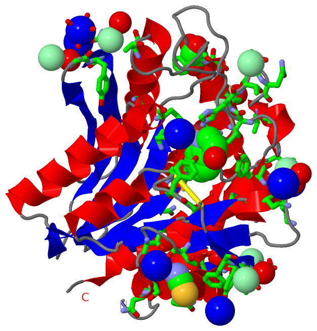
| 01 | AC1 | SOFTWARE | ASP A:117 , GLU A:152 , HIS A:256 , ZN A:302 , HQY A:303 | BINDING SITE FOR RESIDUE ZN A 301 |
| 02 | AC2 | SOFTWARE | HIS A:97 , ASP A:117 , GLU A:152 , ASP A:179 , ZN A:301 , HQY A:303 | BINDING SITE FOR RESIDUE ZN A 302 |
| 03 | AC3 | SOFTWARE | HIS A:97 , ASP A:117 , GLU A:152 , ASP A:179 , GLY A:224 , TYR A:225 , CYS A:227 , TYR A:251 , ILE A:255 , HIS A:256 , ZN A:301 , ZN A:302 , HOH A:451 , HOH A:508 | BINDING SITE FOR RESIDUE HQY A 303 |
| 04 | AC4 | SOFTWARE | THR A:194 , THR A:197 , HOH A:468 , HOH A:600 , HOH A:625 | BINDING SITE FOR RESIDUE NA A 304 |
| 05 | AC5 | SOFTWARE | SER A:51 , ASN A:74 , NA A:312 , HOH A:496 , HOH A:498 , HOH A:501 , HOH A:623 | BINDING SITE FOR RESIDUE NA A 305 |
| 06 | AC6 | SOFTWARE | TYR A:218 , HOH A:441 , HOH A:455 , HOH A:471 , HOH A:510 , HOH A:620 | BINDING SITE FOR RESIDUE NA A 306 |
| 07 | AC7 | SOFTWARE | ASN A:249 , HOH A:357 , HOH A:442 , HOH A:515 , HOH A:525 , HOH A:561 | BINDING SITE FOR RESIDUE NA A 307 |
| 08 | AC8 | SOFTWARE | THR A:268 , SER A:270 , HOH A:355 , HOH A:567 , HOH A:609 , HOH A:627 | BINDING SITE FOR RESIDUE NA A 308 |
| 09 | AC9 | SOFTWARE | THR A:11 , HOH A:352 , HOH A:353 , HOH A:473 , HOH A:619 | BINDING SITE FOR RESIDUE NA A 309 |
| 10 | BC1 | SOFTWARE | GLU A:152 , HOH A:451 , HOH A:479 , HOH A:534 , HOH A:535 | BINDING SITE FOR RESIDUE NA A 310 |
| 11 | BC2 | SOFTWARE | HOH A:329 , HOH A:333 , HOH A:359 , HOH A:378 , HOH A:416 , HOH A:579 | BINDING SITE FOR RESIDUE NA A 311 |
| 12 | BC3 | SOFTWARE | ASN A:74 , NA A:305 , HOH A:501 , HOH A:549 , HOH A:565 , HOH A:616 , HOH A:691 | BINDING SITE FOR RESIDUE NA A 312 |
| 13 | BC4 | SOFTWARE | LEU A:213 , PRO A:214 , SER A:215 | BINDING SITE FOR RESIDUE CL A 313 |
| 14 | BC5 | SOFTWARE | ALA A:19 , LYS A:273 , HOH A:590 | BINDING SITE FOR RESIDUE CL A 314 |
| 15 | BC6 | SOFTWARE | THR A:257 , THR A:258 , GLN A:259 | BINDING SITE FOR RESIDUE CL A 315 |
| 16 | BC7 | SOFTWARE | THR A:38 , TYR A:73 , ASN A:74 , HOH A:397 , HOH A:549 | BINDING SITE FOR RESIDUE CL A 316 |
| 17 | BC8 | SOFTWARE | GLY A:72 , HOH A:466 | BINDING SITE FOR RESIDUE CL A 317 |
| 18 | BC9 | SOFTWARE | GLN A:6 , ALA A:8 , THR A:9 , HOH A:516 | BINDING SITE FOR RESIDUE CL A 318 |
| 19 | CC1 | SOFTWARE | GLY A:24 , SER A:27 , SER A:28 , ASN A:200 , HOH A:493 | BINDING SITE FOR RESIDUE CL A 319 |
| 20 | CC2 | SOFTWARE | LYS A:184 , HOH A:366 , HOH A:387 | BINDING SITE FOR RESIDUE CL A 320 |
| 21 | CC3 | SOFTWARE | GLY A:219 , PHE A:220 , HOH A:345 | BINDING SITE FOR RESIDUE CL A 321 |
| 22 | CC4 | SOFTWARE | THR A:194 , SER A:199 , THR A:202 , GLN A:203 , LEU A:262 , HOH A:625 | BINDING SITE FOR RESIDUE SCN A 322 |
| 23 | CC5 | SOFTWARE | TRP A:13 , GLN A:16 , ASN A:135 , HOH A:352 , HOH A:539 | BINDING SITE FOR RESIDUE SCN A 323 |
| 24 | CC6 | SOFTWARE | SER A:28 , SER A:31 , PHE A:32 , HOH A:411 | BINDING SITE FOR RESIDUE GOL A 324 |
 Description