|
|
|
|
 Description
|
|
Compounds
|
||||||||||||||||||||||||||||||||||||
Chains, Units
Summary Information (see also Sequences/Alignments below) |
Ligands, Modified Residues, Ions (2, 3)| Asymmetric/Biological Unit (2, 3) |
Sites (3, 3)
Asymmetric Unit (3, 3)
|
SS Bonds (1, 1)
Asymmetric/Biological Unit
|
||||||||
Cis Peptide Bonds (0, 0)| (no "Cis Peptide Bond" information available for 2PRQ) |
SAPs(SNPs)/Variants (0, 0)| (no "SAP(SNP)/Variant" information available for 2PRQ) |
PROSITE Motifs (0, 0)| (no "PROSITE Motif" information available for 2PRQ) |
Exons (0, 0)| (no "Exon" information available for 2PRQ) |
Sequences/Alignments
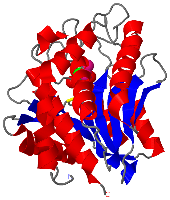
Asymmetric/Biological UnitChain A from PDB Type:PROTEIN Length:291 aligned with AMPX_VIBPR | Q01693 from UniProtKB/Swiss-Prot Length:504 Alignment length:291 116 126 136 146 156 166 176 186 196 206 216 226 236 246 256 266 276 286 296 306 316 326 336 346 356 366 376 386 396 AMPX_VIBPR 107 MPPITQQATVTAWLPQVDASQITGTISSLESFTNRFYTTTSGAQASDWIASEWQALSASLPNASVKQVSHSGYNQKSVVMTITGSEAPDEWIVIGGHLDSTIGSHTNEQSVAPGADDDASGIAAVTEVIRVLSENNFQPKRSIAFMAYAAEEVGLRGSQDLANQYKSEGKNVVSALQLDMTNYKGSAQDVVFITDYTDSNFTQYLTQLMDEYLPSLTYGFDTCGYACSDHASWHNAGYPAAMPFESKFNDYNPRIHTTQDTLANSDPTGSHAKKFTQLGLAYAIEMGSATG 397 SCOP domains d2prqa_ A: Aminopeptidase SCOP domains CATH domains 2prqA00 A:1-291 Zn peptidases CATH domains Pfam domains -----------------------------------------------------------------------------------------Peptidase_M28-2prqA01 A:90-271 -------------------- Pfam domains SAPs(SNPs) --------------------------------------------------------------------------------------------------------------------------------------------------------------------------------------------------------------------------------------------------------------------------------------------------- SAPs(SNPs) PROSITE --------------------------------------------------------------------------------------------------------------------------------------------------------------------------------------------------------------------------------------------------------------------------------------------------- PROSITE Transcript --------------------------------------------------------------------------------------------------------------------------------------------------------------------------------------------------------------------------------------------------------------------------------------------------- Transcript 2prq A 1 MPPITQQATVTAWLPQVDASQITGTISSLESFTNRFYTTTSGAQASDWIASEWQALSASLPNASVKQVSHSGYNQKSVVMTITGSEAPDEWIVIGGHLDSTIGSHTNEQSVAPGADDDASGIAAVTEVIRVLSENNFQPKRSIAFMAYAAEEVGLRGSQDLANQYKSEGKNVVSALQLDMTNYKGSAQDVVFITDYTDSNFTQYLTQLMDEYLPSLTYGFDTCGYACSDHASWHNAGYPAAMPFESKFNDYNPRIHTTQDTLANSDPTGSHAKKFTQLGLAYAIEMGSATG 291 10 20 30 40 50 60 70 80 90 100 110 120 130 140 150 160 170 180 190 200 210 220 230 240 250 260 270 280 290
|
||||||||||||||||||||
SCOP Domains (1, 1)
Asymmetric/Biological Unit
 |
CATH Domains (1, 1)
Asymmetric/Biological Unit
|
Pfam Domains (1, 1)| Asymmetric/Biological Unit |
Gene Ontology (6, 6)|
Asymmetric/Biological Unit(hide GO term definitions) Chain A (AMPX_VIBPR | Q01693)
|
||||||||||||||||||||||||||||||||||||||||||||||||||||||
Interactive Views
|
|||||||||||||||||||||||||||||||||||||||||||||||||||||||||||||||||||||||||||||||||||||||||||||||||||||||||||||||||||||||||||||||||||||||||||
Still Images
|
||||||||||||||||
Databases
|
||||||||||||||||||||||||||||||||||||||||||||||||||||||||||||||||||||||||||||||||||||||||||||||||||||||||||||||||||||||||||||||||||||||||||||||||||||||||||||||||
Analysis Tools
|
|||||||||||||||||||||||||||||||||||||||||||||||||||||||||||||
Entries Sharing at Least One Protein Chain (UniProt ID)
Related Entries Specified in the PDB File
|
|