Show PDB file:   
         Plain Text   HTML   (compressed file size)
QuickSearch:   
by PDB,NDB,UniProt,PROSITE Code or Search Term(s)  
(-)Asymmetric Unit
(-)Asym. Unit - sites
(-)Biological Unit 1
(-)Biol. Unit 1 - sites
collapse expand < >
Image Asymmetric Unit
Asymmetric Unit  (Jmol Viewer)
Image Asym. Unit - sites
Asym. Unit - sites  (Jmol Viewer)
Image Biological Unit 1
Biological Unit 1  (Jmol Viewer)
Image Biol. Unit 1 - sites
Biol. Unit 1 - sites  (Jmol Viewer)

(-) Description

Title :  STRUCTURE OF RECOMBINATION MEDIATOR PROTEIN RECRK21G MUTANT
 
Authors :  Q. Tang, X. X. Yan, D. C. Liang
Date :  06 Jan 12  (Deposition) - 19 Dec 12  (Release) - 19 Dec 12  (Revision)
Method :  X-RAY DIFFRACTION
Resolution :  2.80
Chains :  Asym. Unit :  A
Biol. Unit 1:  A  (2x)
Keywords :  Zinc Finger, Dna Repair, Dna Binding, Recombination (Keyword Search: [Gene Ontology, PubMed, Web (Google))
 
Reference :  Q. Tang, P. Gao, Y. P. Liu, A. Gao, X. M. An, S. Liu, X. X. Yan, D. C. Liang
Recor Complex Including Recr N-N Dimer And Reco Monomer Displays A High Affinity For Ssdna
Nucleic Acids Res. V. 40 11115 2012
PubMed-ID: 23019218  |  Reference-DOI: 10.1093/NAR/GKS889

(-) Compounds

Molecule 1 - RECOMBINATION PROTEIN RECR
    ChainsA
    EngineeredYES
    Expression SystemESCHERICHIA COLI
    Expression System Taxid562
    GeneRECR
    MutationYES
    Organism ScientificTHERMOANAEROBACTER TENGCONGENSIS
    Organism Taxid273068
    StrainMB4
    SynonymRECR RECOMBINATIONAL DNA REPAIR PROTEIN

 Structural Features

(-) Chains, Units

  1
Asymmetric Unit A
Biological Unit 1 (2x)A

Summary Information (see also Sequences/Alignments below)

(-) Ligands, Modified Residues, Ions  (1, 1)

Asymmetric Unit (1, 1)
No.NameCountTypeFull Name
1ZN1Ligand/IonZINC ION
Biological Unit 1 (0, 0)
No.NameCountTypeFull Name
1ZN-1Ligand/IonZINC ION

(-) Sites  (1, 1)

Asymmetric Unit (1, 1)
No.NameEvidenceResiduesDescription
1AC1SOFTWARECYS A:55 , CYS A:58 , CYS A:67 , CYS A:70BINDING SITE FOR RESIDUE ZN A 201

(-) SS Bonds  (0, 0)

(no "SS Bond" information available for 3VDU)

(-) Cis Peptide Bonds  (0, 0)

(no "Cis Peptide Bond" information available for 3VDU)

 Sequence-Structure Mapping

(-) SAPs(SNPs)/Variants  (0, 0)

(no "SAP(SNP)/Variant" information available for 3VDU)

(-) PROSITE Motifs  (2, 2)

Asymmetric Unit (2, 2)
 PROSITEUniProtKBPDB
No.IDACDescriptionIDLocationCountLocation
1RECRPS01300 RecR protein signature.RECR_CALS458-79  1A:55-76
2TOPRIMPS50880 Toprim domain profile.RECR_CALS481-176  1A:78-173
Biological Unit 1 (2, 4)
 PROSITEUniProtKBPDB
No.IDACDescriptionIDLocationCountLocation
1RECRPS01300 RecR protein signature.RECR_CALS458-79  2A:55-76
2TOPRIMPS50880 Toprim domain profile.RECR_CALS481-176  2A:78-173

(-) Exons   (0, 0)

(no "Exon" information available for 3VDU)

(-) Sequences/Alignments

Asymmetric Unit
   Reformat: Number of residues per line =  ('0' or empty: single-line sequence representation)
  Number of residues per labelling interval =   
  UniProt sequence: complete  aligned part    
   Show mapping: SCOP domains CATH domains Pfam domains Secondary structure (by author)
SAPs(SNPs) PROSITE motifs Exons
(details for a mapped element are shown in a popup box when the mouse pointer rests over it)
Chain A from PDB  Type:PROTEIN  Length:197
 aligned with RECR_CALS4 | Q8RDI4 from UniProtKB/Swiss-Prot  Length:199

    Alignment length:197
                                    12        22        32        42        52        62        72        82        92       102       112       122       132       142       152       162       172       182       192       
           RECR_CALS4     3 YYSTSVAKLIEELSKLPGIGPKTAQRLAFFIINMPLDEVRSLSQAIIEAKEKLRYCKICFNITDKEVCDICSDENRDHSTICVVSHPMDVVAMEKVKEYKGVYHVLHGVISPIEGVGPEDIRIKELLERVRDGSVKEVILATNPDIEGEATAMYIAKLLKPFGVKVTRIAHGIPVGGDLEYTDVVTLSKALEGRREV 199
               SCOP domains d3vdua_ A: Recombination protein RecR                                                                                                                                                                 SCOP domains
               CATH domains ----------------------------------------------------------------------------------------------------------------------------------------------------------------------------------------------------- CATH domains
               Pfam domains ----------------------------------------------------------------------------------------------------------------------------------------------------------------------------------------------------- Pfam domains
         Sec.struct. author ..hhhhhhhhhhhhh....hhhhhhhhhhhhhhhhhhhhhhhhhhhhhhhhhhee......ee.............eeeeeeee..hhhhhhhhh.....eeeee.................hhhhhhhhhh....eeee.........hhhhhhhhhhh....eeee..............hhhhhhhhhhh.... Sec.struct. author
                 SAPs(SNPs) ----------------------------------------------------------------------------------------------------------------------------------------------------------------------------------------------------- SAPs(SNPs)
                    PROSITE -------------------------------------------------------RECR  PDB: A:55-76    -TOPRIM  PDB: A:78-173 UniProt: 81-176                                                           ----------------------- PROSITE
                 Transcript ----------------------------------------------------------------------------------------------------------------------------------------------------------------------------------------------------- Transcript
                 3vdu A   0 YYSTSVAKLIEELSKLPGIGPGTAQRLAFFIINMPLDEVRSLSQAIIEAKEKLRYCKICFNITDKEVCDICSDENRDHSTICVVSHPMDVVAMEKVKEYKGVYHVLHGVISPIEGVGPEDIRIKELLERVRDGSVKEVILATNPDIEGEATAMYIAKLLKPFGVKVTRIAHGIPVGGDLEYTDVVTLSKALEGRREV 196
                                     9        19        29        39        49        59        69        79        89        99       109       119       129       139       149       159       169       179       189       

   Legend:   → Mismatch (orange background)
  - → Gap (green background, '-', border residues have a numbering label)
    → Modified Residue (blue background, lower-case, 'x' indicates undefined single-letter code, labelled with number + name)
  x → Chemical Group (purple background, 'x', labelled with number + name, e.g. ACE or NH2)
  extra numbering lines below/above indicate numbering irregularities and modified residue names etc., number ends below/above '|'

 Classification and Annotation

(-) SCOP Domains  (1, 1)

Asymmetric Unit

(-) CATH Domains  (0, 0)

(no "CATH Domain" information available for 3VDU)

(-) Pfam Domains  (0, 0)

(no "Pfam Domain" information available for 3VDU)

(-) Gene Ontology  (5, 5)

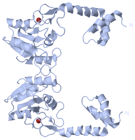
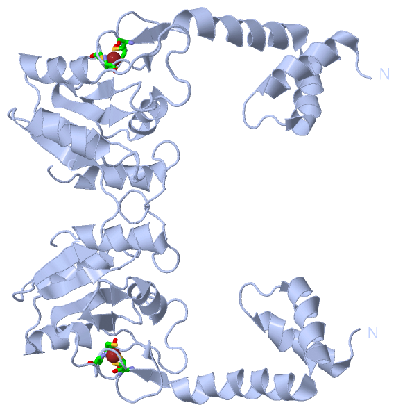
Asymmetric Unit(hide GO term definitions)
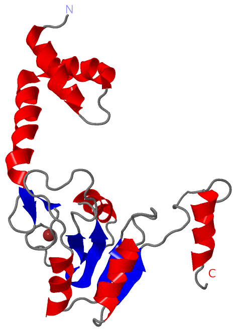
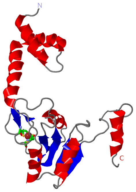
Chain A   (RECR_CALS4 | Q8RDI4)
molecular function
    GO:0003677    DNA binding    Any molecular function by which a gene product interacts selectively and non-covalently with DNA (deoxyribonucleic acid).
    GO:0046872    metal ion binding    Interacting selectively and non-covalently with any metal ion.
biological process
    GO:0006310    DNA recombination    Any process in which a new genotype is formed by reassortment of genes resulting in gene combinations different from those that were present in the parents. In eukaryotes genetic recombination can occur by chromosome assortment, intrachromosomal recombination, or nonreciprocal interchromosomal recombination. Intrachromosomal recombination occurs by crossing over. In bacteria it may occur by genetic transformation, conjugation, transduction, or F-duction.
    GO:0006281    DNA repair    The process of restoring DNA after damage. Genomes are subject to damage by chemical and physical agents in the environment (e.g. UV and ionizing radiations, chemical mutagens, fungal and bacterial toxins, etc.) and by free radicals or alkylating agents endogenously generated in metabolism. DNA is also damaged because of errors during its replication. A variety of different DNA repair pathways have been reported that include direct reversal, base excision repair, nucleotide excision repair, photoreactivation, bypass, double-strand break repair pathway, and mismatch repair pathway.
    GO:0006974    cellular response to DNA damage stimulus    Any process that results in a change in state or activity of a cell (in terms of movement, secretion, enzyme production, gene expression, etc.) as a result of a stimulus indicating damage to its DNA from environmental insults or errors during metabolism.

 Visualization

(-) Interactive Views

Asymmetric Unit
  Complete Structure
    Jena3D(integrated viewing of ligand, site, SAP, PROSITE, SCOP information)
    WebMol | AstexViewer[tm]@PDBe
(Java Applets, require no local installation except for Java; loading may be slow)
    STRAP
(Java WebStart application, automatic local installation, requires Java; full application with system access!)
    RasMol
(require local installation)
    Molscript (VRML)
(requires installation of a VRML viewer; select preferred view via VRML and generate a mono or stereo PDF format file)
 
  Ligands, Modified Residues, Ions
    ZN  [ RasMol | Jena3D ]  +environment [ RasMol | Jena3D ]
 
  Sites
    AC1  [ RasMol ]  +environment [ RasMol ]
 
  Cis Peptide Bonds
(no "Cis Peptide Bonds" information available for 3vdu)
 
Biological Unit
  Complete Structure
    Biological Unit 1  [ Jena3D ]

(-) Still Images

Jmol
  protein: cartoon or spacefill or dots and stick; nucleic acid: cartoon and stick; ligands: spacefill; active site: stick
Molscript
  protein, nucleic acid: cartoon; ligands: spacefill; active site: ball and stick

 Databases and Analysis Tools

(-) Databases

Access by PDB/NDB ID
  3vdu
    Family and Domain InformationProDom | SYSTERS
    General Structural InformationGlycoscienceDB | MMDB | NDB | OCA | PDB | PDBe | PDBj | PDBsum | PDBWiki | PQS | PROTEOPEDIA
    Orientation in MembranesOPM
    Protein SurfaceSURFACE
    Secondary StructureDSSP (structure derived) | HSSP (homology derived)
    Structural GenomicsGeneCensus
    Structural NeighboursCE | VAST
    Structure ClassificationCATH | Dali | SCOP
    Validation and Original DataBMRB Data View | BMRB Restraints Grid | EDS | PROCHECK | RECOORD | WHAT_CHECK
 
Access by UniProt ID/Accession number
  RECR_CALS4 | Q8RDI4
    Comparative Protein Structure ModelsModBase
    Genomic InformationEnsembl
    Protein-protein InteractionDIP
    Sequence, Family and Domain InformationInterPro | Pfam | SMART | UniProtKB/SwissProt
 
Access by Enzyme Classificator   (EC Number)
  (no 'Enzyme Classificator' available)
    General Enzyme InformationBRENDA | EC-PDB | Enzyme | IntEnz
    PathwayKEGG | MetaCyc
 
Access by Disease Identifier   (MIM ID)
  (no 'MIM ID' available)
    Disease InformationOMIM
 
Access by GenAge ID
  (no 'GenAge ID' available)
    Age Related InformationGenAge

(-) Analysis Tools

Access by PDB/NDB ID
    Domain InformationXDom
    Interatomic Contacts of Structural UnitsCSU
    Ligand-protein ContactsLPC
    Protein CavitiescastP
    Sequence and Secondary StructurePDBCartoon
    Structure AlignmentSTRAP(Java WebStart application, automatic local installation, requires Java; full application with system access!)
    Structure and Sequence BrowserSTING
 
Access by UniProt ID/Accession number
  RECR_CALS4 | Q8RDI4
    Protein Disorder PredictionDisEMBL | FoldIndex | GLOBPLOT (for more information see DisProt)

 Related Entries

(-) Entries Sharing at Least One Protein Chain (UniProt ID)

UniProtKB/Swiss-Prot
        RECR_CALS4 | Q8RDI43vdp 3ve5 4o6o 4o6p

(-) Related Entries Specified in the PDB File

3vdp THE SAME PROTEIN, APO-FORM
3vds THE SINGLE POINT MUTANT TTERECR C55G OF SAME PROTEIN
3vdt THE DOUBLE POINTS MUTANT TTERECR C58G/C70G OF SAME PROTEIN
3ve5 THE DELETE MUTANT OF RECR PROTEIN