|
|
|
|
 Description
|
|
Compounds
|
||||||||||||||||||||||||||||||||||||||||||||
Chains, Units
Summary Information (see also Sequences/Alignments below) |
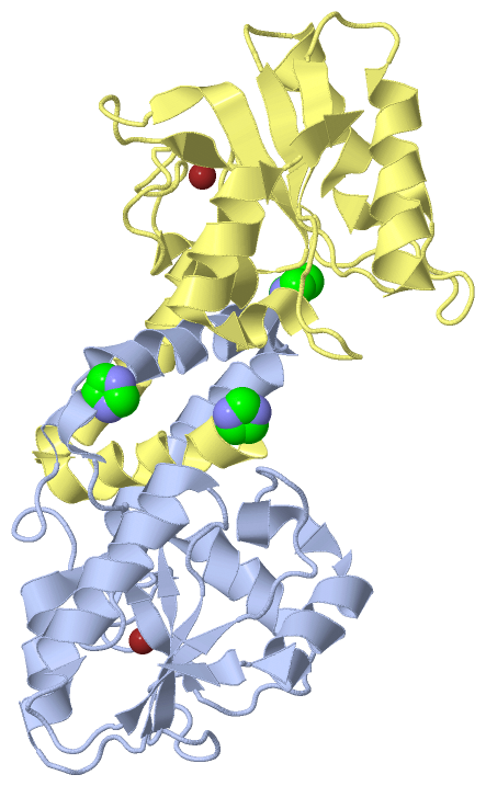
Ligands, Modified Residues, Ions (2, 5)| Asymmetric/Biological Unit (2, 5) |
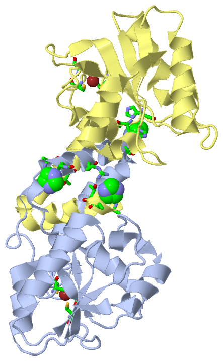
Sites (5, 5)
Asymmetric Unit (5, 5)
|
SS Bonds (0, 0)| (no "SS Bond" information available for 3VE5) |
Cis Peptide Bonds (2, 2)
Asymmetric/Biological Unit
|
||||||||||||
SAPs(SNPs)/Variants (0, 0)| (no "SAP(SNP)/Variant" information available for 3VE5) |
PROSITE Motifs (2, 4)
Asymmetric/Biological Unit (2, 4)
|
||||||||||||||||||||||||||||||||
Exons (0, 0)| (no "Exon" information available for 3VE5) |
Sequences/Alignments
Asymmetric/Biological UnitChain A from PDB Type:PROTEIN Length:181 aligned with RECR_CALS4 | Q8RDI4 from UniProtKB/Swiss-Prot Length:199 Alignment length:181 28 38 48 58 68 78 88 98 108 118 128 138 148 158 168 178 188 198 RECR_CALS4 19 PGIGPKTAQRLAFFIINMPLDEVRSLSQAIIEAKEKLRYCKICFNITDKEVCDICSDENRDHSTICVVSHPMDVVAMEKVKEYKGVYHVLHGVISPIEGVGPEDIRIKELLERVRDGSVKEVILATNPDIEGEATAMYIAKLLKPFGVKVTRIAHGIPVGGDLEYTDVVTLSKALEGRREV 199 SCOP domains ------------------------------------------------------------------------------------------------------------------------------------------------------------------------------------- SCOP domains CATH domains ------------------------------------------------------------------------------------------------------------------------------------------------------------------------------------- CATH domains Pfam domains ------------------------------------------------------------------------------------------------------------------------------------------------------------------------------------- Pfam domains SAPs(SNPs) ------------------------------------------------------------------------------------------------------------------------------------------------------------------------------------- SAPs(SNPs) PROSITE ---------------------------------------RECR PDB: A:55-76 -TOPRIM PDB: A:78-173 UniProt: 81-176 ----------------------- PROSITE Transcript ------------------------------------------------------------------------------------------------------------------------------------------------------------------------------------- Transcript 3ve5 A 16 PGIGPKTAQRLAFFIINMPLDEVRSLSQAIIEAKEKLRYCKICFNITDKEVCDICSDENRDHSTICVVSHPMDVVAMEKVKEYKGVYHVLHGVISPIEGVGPEDIRIKELLERVRDGSVKEVILATNPDIEGEATAMYIAKLLKPFGVKVTRIAHGIPVGGDLEYTDVVTLSKALEGRREV 196 25 35 45 55 65 75 85 95 105 115 125 135 145 155 165 175 185 195 Chain D from PDB Type:PROTEIN Length:181 aligned with RECR_CALS4 | Q8RDI4 from UniProtKB/Swiss-Prot Length:199 Alignment length:181 28 38 48 58 68 78 88 98 108 118 128 138 148 158 168 178 188 198 RECR_CALS4 19 PGIGPKTAQRLAFFIINMPLDEVRSLSQAIIEAKEKLRYCKICFNITDKEVCDICSDENRDHSTICVVSHPMDVVAMEKVKEYKGVYHVLHGVISPIEGVGPEDIRIKELLERVRDGSVKEVILATNPDIEGEATAMYIAKLLKPFGVKVTRIAHGIPVGGDLEYTDVVTLSKALEGRREV 199 SCOP domains ------------------------------------------------------------------------------------------------------------------------------------------------------------------------------------- SCOP domains CATH domains ------------------------------------------------------------------------------------------------------------------------------------------------------------------------------------- CATH domains Pfam domains ------------------------------------------------------------------------------------------------------------------------------------------------------------------------------------- Pfam domains SAPs(SNPs) ------------------------------------------------------------------------------------------------------------------------------------------------------------------------------------- SAPs(SNPs) PROSITE ---------------------------------------RECR PDB: D:55-76 -TOPRIM PDB: D:78-173 UniProt: 81-176 ----------------------- PROSITE Transcript ------------------------------------------------------------------------------------------------------------------------------------------------------------------------------------- Transcript 3ve5 D 16 PGIGPKTAQRLAFFIINMPLDEVRSLSQAIIEAKEKLRYCKICFNITDKEVCDICSDENRDHSTICVVSHPMDVVAMEKVKEYKGVYHVLHGVISPIEGVGPEDIRIKELLERVRDGSVKEVILATNPDIEGEATAMYIAKLLKPFGVKVTRIAHGIPVGGDLEYTDVVTLSKALEGRREV 196 25 35 45 55 65 75 85 95 105 115 125 135 145 155 165 175 185 195
|
||||||||||||||||||||
SCOP Domains (0, 0)| (no "SCOP Domain" information available for 3VE5) |
CATH Domains (0, 0)| (no "CATH Domain" information available for 3VE5) |
Pfam Domains (0, 0)| (no "Pfam Domain" information available for 3VE5) |
Gene Ontology (5, 5)|
Asymmetric/Biological Unit(hide GO term definitions) Chain A,D (RECR_CALS4 | Q8RDI4)
|
||||||||||||||||||||||||||||||||||||||||||
Interactive Views
|
|||||||||||||||||||||||||||||||||||||||||||||||||||||||||||||||||||||||||||||||||||||||||||||||||||||||||||||||||||||||||||||||||||||||||||||||||||||||||||||||||
Still Images
|
||||||||||||||||
Databases
|
||||||||||||||||||||||||||||||||||||||||||||||||||||||||||||||||||||||||||||||||||||||||||||||||||||||||||||||||||||||||||||||||||||||||||||||||||||||||||||||||
Analysis Tools
|
|||||||||||||||||||||||||||||||||||||||||||||||||||||||||||||
Entries Sharing at Least One Protein Chain (UniProt ID)
Related Entries Specified in the PDB File
|
|