|
|
|
|
 Description
|
|
Compounds
|
||||||||||||||||||||||||||||||||||||||||||||
Chains, Units
Summary Information (see also Sequences/Alignments below) |
Ligands, Modified Residues, Ions (0, 0)| (no "Ligand,Modified Residues,Ions" information available for 3VDJ) |
Sites (0, 0)| (no "Site" information available for 3VDJ) |
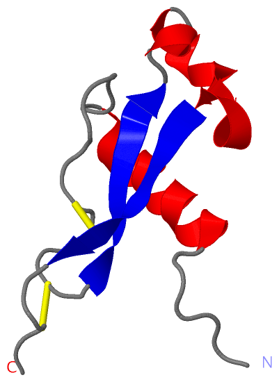
SS Bonds (2, 2)
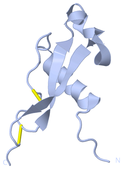
Asymmetric Unit
|
||||||||||||
Cis Peptide Bonds (0, 0)| (no "Cis Peptide Bond" information available for 3VDJ) |
SAPs(SNPs)/Variants (0, 0)| (no "SAP(SNP)/Variant" information available for 3VDJ) |
PROSITE Motifs (0, 0)| (no "PROSITE Motif" information available for 3VDJ) |
Exons (0, 0)| (no "Exon" information available for 3VDJ) |
Sequences/Alignments
Asymmetric UnitChain A from PDB Type:PROTEIN Length:71 aligned with Q7K740_PLAF7 | Q7K740 from UniProtKB/TrEMBL Length:397 Alignment length:85 301 311 321 331 341 351 361 371 Q7K740_PLAF7 292 NVDENANANSAVKNNNNEEPSDKHIKEYLNKIQNSLSTEWSPCSVTCGNGIQVRIKPGSANKPKDELDYANDIEKKICKMEKCSS 376 SCOP domains ------------------------------------------------------------------------------------- SCOP domains CATH domains ------------------------------------------------------------------------------------- CATH domains Pfam domains ------------------------------------------------------------------------------------- Pfam domains SAPs(SNPs) ------------------------------------------------------------------------------------- SAPs(SNPs) PROSITE ------------------------------------------------------------------------------------- PROSITE Transcript ------------------------------------------------------------------------------------- Transcript 3vdj A 306 YVEF--------------EPSDKHIKEYLNKIQNSLSTEWSPCSVTCGNGIQVRIKPGSANKPKDELDYANDIEKKICKMEKCPH 376 | - 311 321 331 341 351 361 371 309 310
|
||||||||||||||||||||
SCOP Domains (0, 0)| (no "SCOP Domain" information available for 3VDJ) |
CATH Domains (0, 0)| (no "CATH Domain" information available for 3VDJ) |
Pfam Domains (0, 0)| (no "Pfam Domain" information available for 3VDJ) |
Gene Ontology (7, 7)|
Asymmetric Unit(hide GO term definitions) Chain A (Q7K740_PLAF7 | Q7K740)
|
||||||||||||||||||||||||||||||||||||||||||||||||||||||||||||
Interactive Views
|
|||||||||||||||||||||||||||||||||||||||||||||||||||||||||||||||||||||||||||||||||||||||||||||||||||||||||||||||||||||||||||||||||||||||||||
Still Images
|
||||||||||||||||
Databases
|
||||||||||||||||||||||||||||||||||||||||||||||||||||||||||||||||||||||||||||||||||||||||||||||||||||||||||||||||||||||||||||||||||||||||||||||||||||||||||||||||
Analysis Tools
|
|||||||||||||||||||||||||||||||||||||||||||||||||||||||||||||
Entries Sharing at Least One Protein Chain (UniProt ID)
Related Entries Specified in the PDB File
|
|