|
|
|
|
 Description
|
|
Compounds
|
||||||||||||||||||||||||||||||||||||||||
Chains, Units
Summary Information (see also Sequences/Alignments below) |
Ligands, Modified Residues, Ions (1, 1)
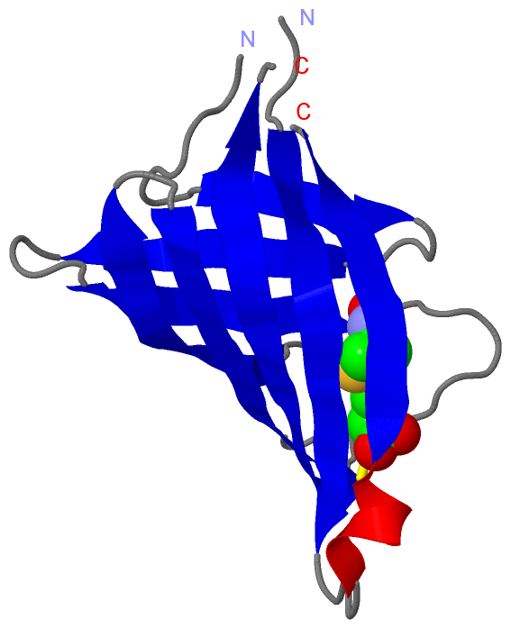
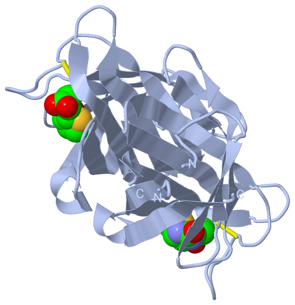
Asymmetric Unit (1, 1)
|
Sites (1, 1)
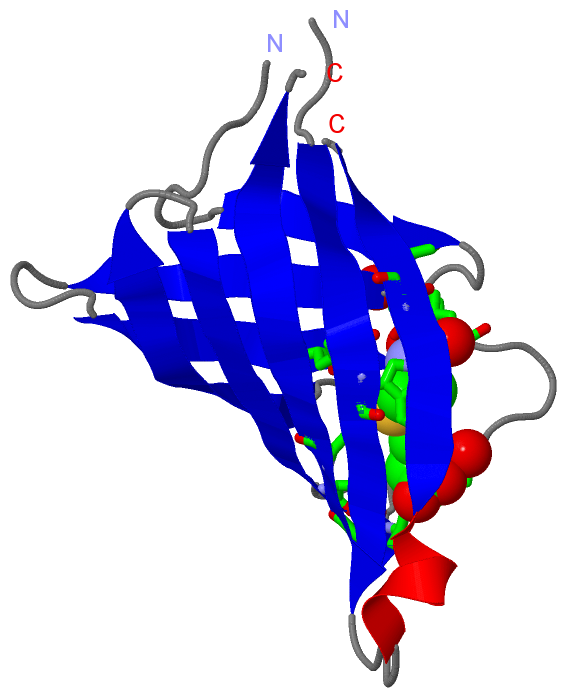
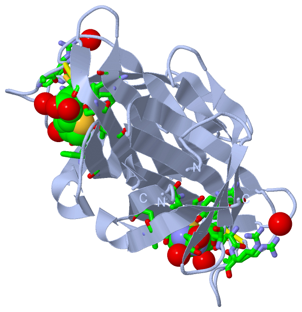
Asymmetric Unit (1, 1)
|
SS Bonds (1, 1)
Asymmetric Unit
|
||||||||
Cis Peptide Bonds (0, 0)| (no "Cis Peptide Bond" information available for 3T2W) |
SAPs(SNPs)/Variants (0, 0)| (no "SAP(SNP)/Variant" information available for 3T2W) |
PROSITE Motifs (0, 0)| (no "PROSITE Motif" information available for 3T2W) |
Exons (0, 0)| (no "Exon" information available for 3T2W) |
Sequences/Alignments
Asymmetric UnitChain A from PDB Type:PROTEIN Length:115 aligned with Q12QS6_SHEDO | Q12QS6 from UniProtKB/TrEMBL Length:172 Alignment length:117 53 63 73 83 93 103 113 123 133 143 153 Q12QS6_SHEDO 44 AQELTAMSAWVNQDGSTLYINSINAQGELTGSYINRAAGFACQNSPYPVNGWVFGTAISFSTKWLNSVESCNSITSWSGFYINTGGQGKISTLWQLVVNGSSSPSQILKGQDVFSQT 160 SCOP domains d3t2wa_ A: automated matches SCOP domains CATH domains --------------------------------------------------------------------------------------------------------------------- CATH domains Pfam domains --------------------------------------------------------------------------------------------------------------------- Pfam domains SAPs(SNPs) --------------------------------------------------------------------------------------------------------------------- SAPs(SNPs) PROSITE --------------------------------------------------------------------------------------------------------------------- PROSITE Transcript --------------------------------------------------------------------------------------------------------------------- Transcript 3t2w A 4 AQELTAMSAWVNQDGSTLYINSINAQGELTGSYINRAAGAACQNSPYPVNGWVFGTAISFSTKWLNSVESCNSITSWSGFYIN--GQGKISTLWQLVVNGSSSPSQILKGQDVFSQT 120 13 23 33 43 53 63 73 83 | | 93 103 113 86 89
|
||||||||||||||||||||
SCOP Domains (1, 1)
Asymmetric Unit
 |
CATH Domains (0, 0)| (no "CATH Domain" information available for 3T2W) |
Pfam Domains (0, 0)| (no "Pfam Domain" information available for 3T2W) |
Gene Ontology (0, 0)|
Asymmetric Unit(hide GO term definitions)
(no "Gene Ontology" information available for 3T2W)
|
Interactive Views
|
||||||||||||||||||||||||||||||||||||||||||||||||||||||||||||||||||||||||||||||||||||||||||||||||||||||||||||||||||||||||||||||||||||||||
Still Images
|
||||||||||||||||
Databases
|
||||||||||||||||||||||||||||||||||||||||||||||||||||||||||||||||||||||||||||||||||||||||||||||||||||||||||||||||||||||||||||||||||||||||||||||||||||||||||||||||
Analysis Tools
|
|||||||||||||||||||||||||||||||||||||||||||||||||||||||||||||
Entries Sharing at Least One Protein Chain (UniProt ID)
Related Entries Specified in the PDB File
|
|