|
|
|
|
 Description
|
|
Compounds
|
||||||||||||||||||||||||||||||||||||
Chains, Units
Summary Information (see also Sequences/Alignments below) |
Ligands, Modified Residues, Ions (5, 6)| Asymmetric Unit (5, 6) Biological Unit 1 (4, 8) |
Sites (6, 6)
Asymmetric Unit (6, 6)
|
SS Bonds (0, 0)| (no "SS Bond" information available for 3SZ0) |
Cis Peptide Bonds (3, 3)
Asymmetric Unit
|
||||||||||||||||
SAPs(SNPs)/Variants (0, 0)| (no "SAP(SNP)/Variant" information available for 3SZ0) |
PROSITE Motifs (0, 0)| (no "PROSITE Motif" information available for 3SZ0) |
Exons (0, 0)| (no "Exon" information available for 3SZ0) |
Sequences/Alignments
Asymmetric UnitChain A from PDB Type:PROTEIN Length:421 aligned with SQRD_ACIF2 | B7JBP8 from UniProtKB/Swiss-Prot Length:434 Alignment length:421 1 | 7 17 27 37 47 57 67 77 87 97 107 117 127 137 147 157 167 177 187 197 207 217 227 237 247 257 267 277 287 297 307 317 327 337 347 357 367 377 387 397 407 417 SQRD_ACIF2 - ---MAHVVILGAGTGGMPAAYEMKEALGSGHEVTLISANDYFQFVPSNPWVGVGWKERDDIAFPIRHYVERKGIHFIAQSAEQIDAEAQNITLADGNTVHYDYLMIATGPKLAFENVPGSDPHEGPVQSICTVDHAERAFAEYQALLREPGPIVIGAMAGASCFGPAYEYAMIVASDLKKRGMRDKIPSFTFITSEPYIGHLGIQGVGDSKGILTKGLKEEGIEAYTNCKVTKVEDNKMYVTQVDEKGETIKEMVLPVKFGMMIPAFKGVPAVAGVEGLCNPGGFVLVDEHQRSKKYANIFAAGIAIAIPPVETTPVPTGAPKTGYMIESMVSAAVHNIKADLEGRKGEQTMGTWNAVCFADMGDRGAAFIALPQLKPRKVDVFAYGRWVHLAKVAFEKYFIRKMKMGVSEPFYEKVLFKM 418 SCOP domains ------------------------------------------------------------------------------------------------------------------------------------------------------------------------------------------------------------------------------------------------------------------------------------------------------------------------------------------------------------------------------------------------------------------------------------- SCOP domains CATH domains ------------------------------------------------------------------------------------------------------------------------------------------------------------------------------------------------------------------------------------------------------------------------------------------------------------------------------------------------------------------------------------------------------------------------------------- CATH domains Pfam domains ------------------------------------------------------------------------------------------------------------------------------------------------------------------------------------------------------------------------------------------------------------------------------------------------------------------------------------------------------------------------------------------------------------------------------------- Pfam domains SAPs(SNPs) ------------------------------------------------------------------------------------------------------------------------------------------------------------------------------------------------------------------------------------------------------------------------------------------------------------------------------------------------------------------------------------------------------------------------------------- SAPs(SNPs) PROSITE ------------------------------------------------------------------------------------------------------------------------------------------------------------------------------------------------------------------------------------------------------------------------------------------------------------------------------------------------------------------------------------------------------------------------------------- PROSITE Transcript ------------------------------------------------------------------------------------------------------------------------------------------------------------------------------------------------------------------------------------------------------------------------------------------------------------------------------------------------------------------------------------------------------------------------------------- Transcript 3sz0 A -2 MRGSAHVVILGAGTGGMPAAYEMKEALGSGHEVTLISANDYFQFVPSNPWVGVGWKERDDIAFPIRHYVERKGIHFIAQSAEQIDAEAQNITLADGNTVHYDYLMIATGPKLAFENVPGSDPHEGPVQSICTVDHAERAFAEYQALLREPGPIVIGAMAGASCFGPAYEYAMIVASDLKKRGMRDKIPSFTFITSEPYIGHLGIQGVGDSKGILTKGLKEEGIEAYTNCKVTKVEDNKMYVTQVDEKGETIKEMVLPVKFGMMIPAFKGVPAVAGVEGLCNPGGFVLVDEHQRSKKYANIFAAGIAIAIPPVETTPVPTGAPKTGYMIESMVSAAVHNIKADLEGRKGEQTMGTWNAVCFADMGDRGAAFIALPQLKPRKVDVFAYGRWVHLAKVAFEKYFIRKMKMGVSEPFYEKVLFKM 418 7 17 27 37 47 57 67 77 87 97 107 117 127 137 147 157 167 177 187 197 207 217 227 237 247 257 267 277 287 297 307 317 327 337 347 357 367 377 387 397 407 417
|
||||||||||||||||||||
SCOP Domains (0, 0)| (no "SCOP Domain" information available for 3SZ0) |
CATH Domains (0, 0)| (no "CATH Domain" information available for 3SZ0) |
Pfam Domains (0, 0)| (no "Pfam Domain" information available for 3SZ0) |
Gene Ontology (5, 5)|
Asymmetric Unit(hide GO term definitions) Chain A (SQRD_ACIF2 | B7JBP8)
|
||||||||||||||||||||||||||||||||||||||||||||||||
Interactive Views
|
||||||||||||||||||||||||||||||||||||||||||||||||||||||||||||||||||||||||||||||||||||||||||||||||||||||||||||||||||||||||||||||||||||||||||||||||||||||||||||||||||||||||||||||||||||||||||||||||||||||||||||||||||||||
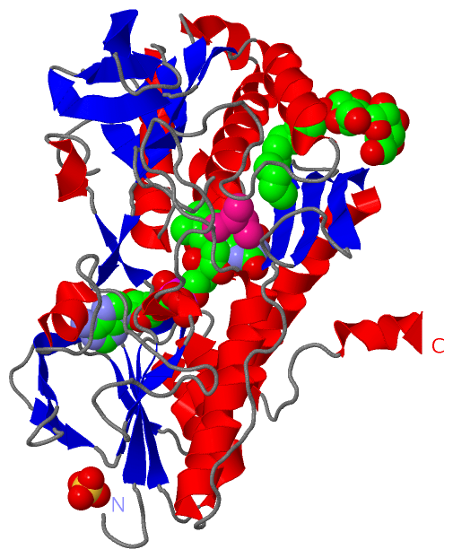
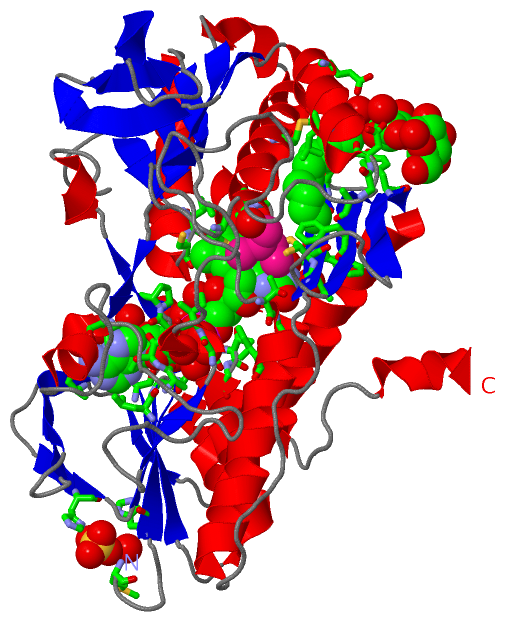
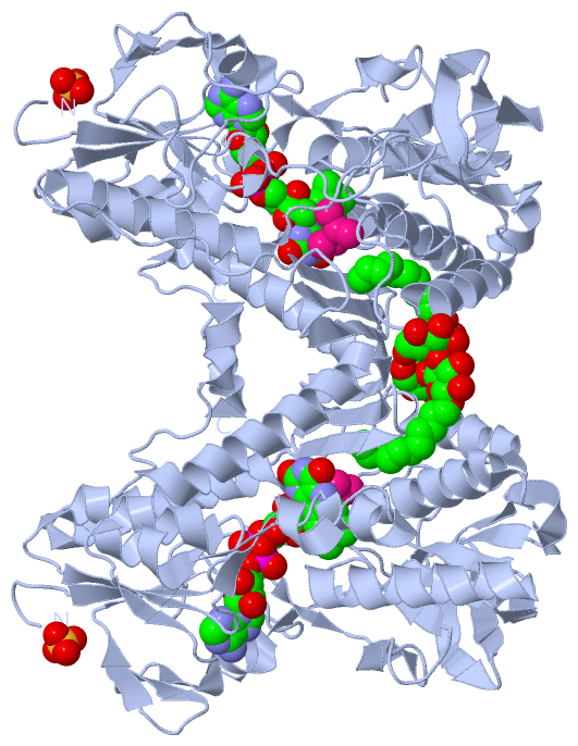
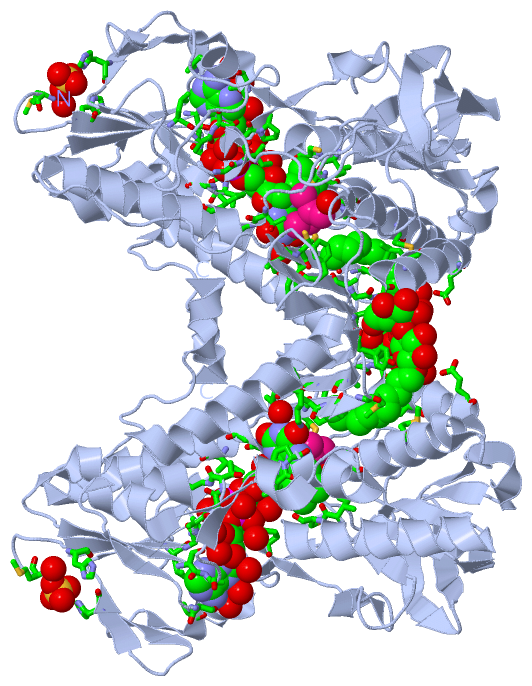
Still Images
|
||||||||||||||||
Databases
|
||||||||||||||||||||||||||||||||||||||||||||||||||||||||||||||||||||||||||||||||||||||||||||||||||||||||||||||||||||||||||||||||||||||||||||||||||||||||||||||||
Analysis Tools
|
|||||||||||||||||||||||||||||||||||||||||||||||||||||||||||||
Entries Sharing at Least One Protein Chain (UniProt ID)
Related Entries Specified in the PDB File
|
|