| molecular function |
|---|
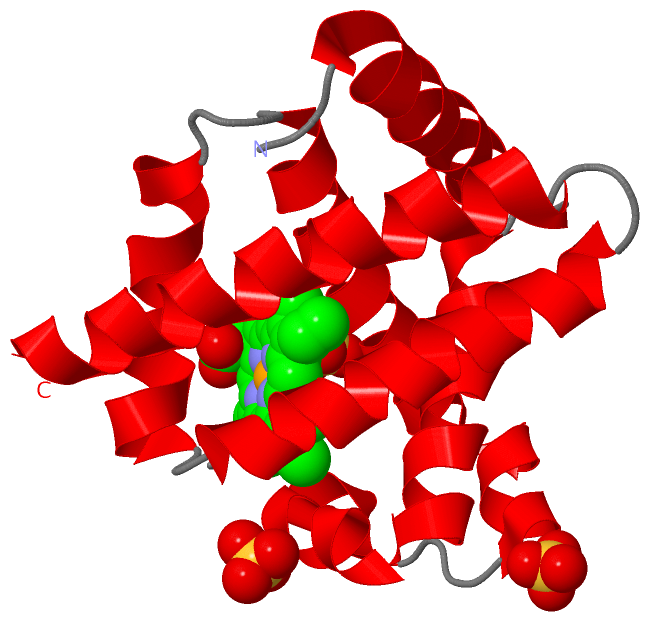
| | GO:0020037 | | heme binding | | Interacting selectively and non-covalently with heme, any compound of iron complexed in a porphyrin (tetrapyrrole) ring. |
| | GO:0046872 | | metal ion binding | | Interacting selectively and non-covalently with any metal ion. |
| | GO:0019825 | | oxygen binding | | Interacting selectively and non-covalently with oxygen (O2). |
| | GO:0005344 | | oxygen transporter activity | | Enables the directed movement of oxygen into, out of or within a cell, or between cells. |
| biological process |
|---|
| | GO:0050873 | | brown fat cell differentiation | | The process in which a relatively unspecialized cell acquires specialized features of a brown adipocyte, an animal connective tissue cell involved in adaptive thermogenesis. Brown adipocytes contain multiple small droplets of triglycerides and a high number of mitochondria. |
| | GO:0043353 | | enucleate erythrocyte differentiation | | The process in which a myeloid precursor cell acquires specialized features of an erythrocyte without a nucleus. An example of this process is found in Mus musculus. |
| | GO:0007507 | | heart development | | The process whose specific outcome is the progression of the heart over time, from its formation to the mature structure. The heart is a hollow, muscular organ, which, by contracting rhythmically, keeps up the circulation of the blood. |
| | GO:0015671 | | oxygen transport | | The directed movement of oxygen (O2) into, out of or within a cell, or between cells, by means of some agent such as a transporter or pore. |
| | GO:0009725 | | response to hormone | | Any process that results in a change in state or activity of a cell or an organism (in terms of movement, secretion, enzyme production, gene expression, etc.) as a result of a hormone stimulus. |
| | GO:0042542 | | response to hydrogen peroxide | | Any process that results in a change in state or activity of a cell or an organism (in terms of movement, secretion, enzyme production, gene expression, etc.) as a result of a hydrogen peroxide (H2O2) stimulus. |
| | GO:0001666 | | response to hypoxia | | Any process that results in a change in state or activity of a cell or an organism (in terms of movement, secretion, enzyme production, gene expression, etc.) as a result of a stimulus indicating lowered oxygen tension. Hypoxia, defined as a decline in O2 levels below normoxic levels of 20.8 - 20.95%, results in metabolic adaptation at both the cellular and organismal level. |
| | GO:0031444 | | slow-twitch skeletal muscle fiber contraction | | A process in which force is generated within slow-twitch skeletal muscle tissue, resulting in a change in muscle geometry. Force generation involves a chemo-mechanical energy conversion step that is carried out by the actin/myosin complex activity, which generates force through ATP hydrolysis. The slow-twitch skeletal muscle is characterized by slow time parameters, low force development and resistance to fatigue. |
| | GO:0006810 | | transport | | The directed movement of substances (such as macromolecules, small molecules, ions) or cellular components (such as complexes and organelles) into, out of or within a cell, or between cells, or within a multicellular organism by means of some agent such as a transporter, pore or motor protein. |
| cellular component |
|---|
| | GO:0070062 | | extracellular exosome | | A vesicle that is released into the extracellular region by fusion of the limiting endosomal membrane of a multivesicular body with the plasma membrane. Extracellular exosomes, also simply called exosomes, have a diameter of about 40-100 nm. |
 Description