|
|
|
|
 Description
|
|
Compounds
|
||||||||||||||||||||||||||||||||||||||||||||||||
Chains, Units
Summary Information (see also Sequences/Alignments below) |
Ligands, Modified Residues, Ions (4, 8)
Asymmetric Unit (4, 8)
|
Sites (8, 8)
Asymmetric Unit (8, 8)
|
SS Bonds (0, 0)| (no "SS Bond" information available for 3OEB) |
Cis Peptide Bonds (0, 0)| (no "Cis Peptide Bond" information available for 3OEB) |
SAPs(SNPs)/Variants (0, 0)| (no "SAP(SNP)/Variant" information available for 3OEB) |
PROSITE Motifs (0, 0)| (no "PROSITE Motif" information available for 3OEB) |
Exons (0, 0)| (no "Exon" information available for 3OEB) |
Sequences/Alignments
Asymmetric UnitChain A from PDB Type:PROTEIN Length:144 aligned with Q9ZA17_9THEO | Q9ZA17 from UniProtKB/TrEMBL Length:1097 Alignment length:144 622 632 642 652 662 672 682 692 702 712 722 732 742 752 Q9ZA17_9THEO 613 GVNMVSNPGFEDGLDSWQDWQQDMSAVPEAAHNGALGLKIGGGKAAGGGQDIPLKPNTTYILGAWAKFDSKPAGTFDVVVQYHLKDANNTYVQHILNFNETDWTYKQLLFTTPDVFGSTPQLALWKGDTSKANLYVDDVYLVEV 756 SCOP domains ------------------------------------------------------------------------------------------------------------------------------------------------ SCOP domains CATH domains ------------------------------------------------------------------------------------------------------------------------------------------------ CATH domains Pfam domains -CBM_4_9-3oebA01 A:2-131 ------------- Pfam domains SAPs(SNPs) ------------------------------------------------------------------------------------------------------------------------------------------------ SAPs(SNPs) PROSITE ------------------------------------------------------------------------------------------------------------------------------------------------ PROSITE Transcript ------------------------------------------------------------------------------------------------------------------------------------------------ Transcript 3oeb A 1 MVNMVSNPGFEDGLDSWQDWQQDMSAVPEAAHNGALGLKIGGGKAAGGGQDIPLKPNTTYILGAWAKFDSKPAGTFDVVVQYHLKDANNTYVQHILNFNETDWTYKQLLFTTPDVFGSTPELALWKGDTSKANLYVDDVYLVEV 144 10 20 30 40 50 60 70 80 90 100 110 120 130 140
|
||||||||||||||||||||
SCOP Domains (0, 0)| (no "SCOP Domain" information available for 3OEB) |
CATH Domains (0, 0)| (no "CATH Domain" information available for 3OEB) |
Pfam Domains (1, 1)
Asymmetric Unit
|
Gene Ontology (6, 6)|
Asymmetric Unit(hide GO term definitions) Chain A (Q9ZA17_9THEO | Q9ZA17)
|
||||||||||||||||||||||||||||||||||||||||||||||||
Interactive Views
|
|||||||||||||||||||||||||||||||||||||||||||||||||||||||||||||||||||||||||||||||||||||||||||||||||||||||||||||||||||||||||||||||||||||||||||||||||||||||||||||||||||||||||||||||||||||||||||||||||||||||||||||||||||
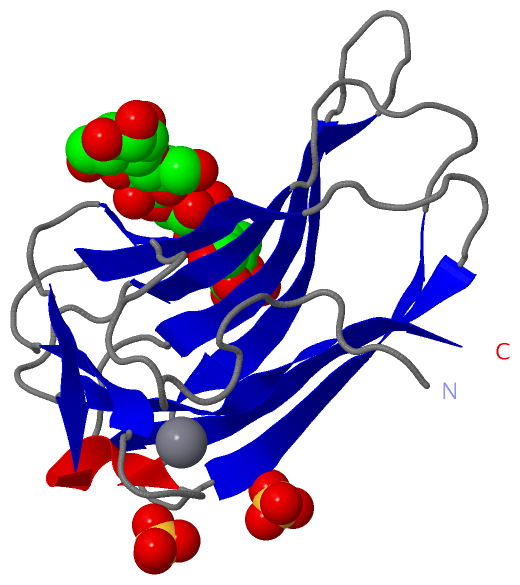
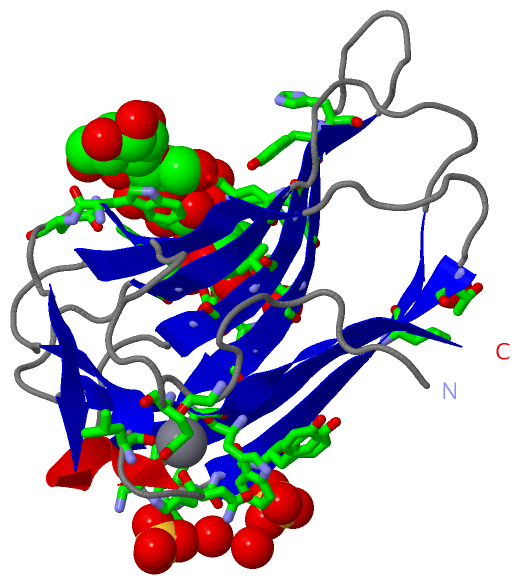
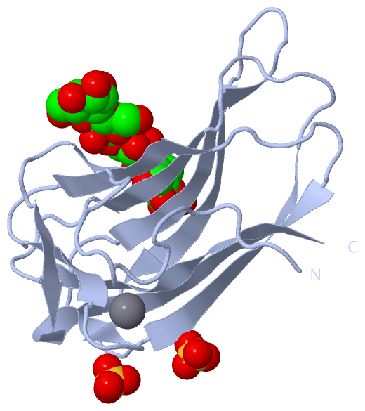
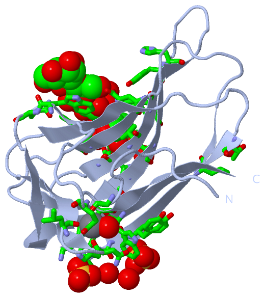
Still Images
|
||||||||||||||||
Databases
|
||||||||||||||||||||||||||||||||||||||||||||||||||||||||||||||||||||||||||||||||||||||||||||||||||||||||||||||||||||||||||||||||||||||||||||||||||||||||||||||||
Analysis Tools
|
|||||||||||||||||||||||||||||||||||||||||||||||||||||||||||||
Entries Sharing at Least One Protein Chain (UniProt ID)
Related Entries Specified in the PDB File
|
|